New ways to image cartilage could help prevent osteoarthritis

Watch this session on ECR Live: Friday, March 8, 16:00–17:30, Room C
Osteoarthritis, a degenerative joint disease, affects a large number of people worldwide. But with the emergence of new MRI techniques, researchers believe they will be able to prevent its development in the near future. Experts will present the latest methods to assess cartilage tissue quality at a very early stage and discuss remaining challenges, in a dedicated New Horizons Session, today at the ECR.
Cartilage is composed of collagen and glycosaminoglycans (GAG), which are responsible for the biomechanical properties of cartilage tissue. An interesting way to image cartilage is to look at the amount of GAG, which decreases at the onset of tissue degeneration, a process which occurs due to ageing or an induced defect, for instance trauma or surgical intervention in the joints. If left untreated, a tissue defect can lead to osteoarthritis. GAGs are known to be among the earliest biomarkers of cartilage degeneration, and if a focal reduction in the amount of GAG can be identified, then therapy to avoid further damage can begin.
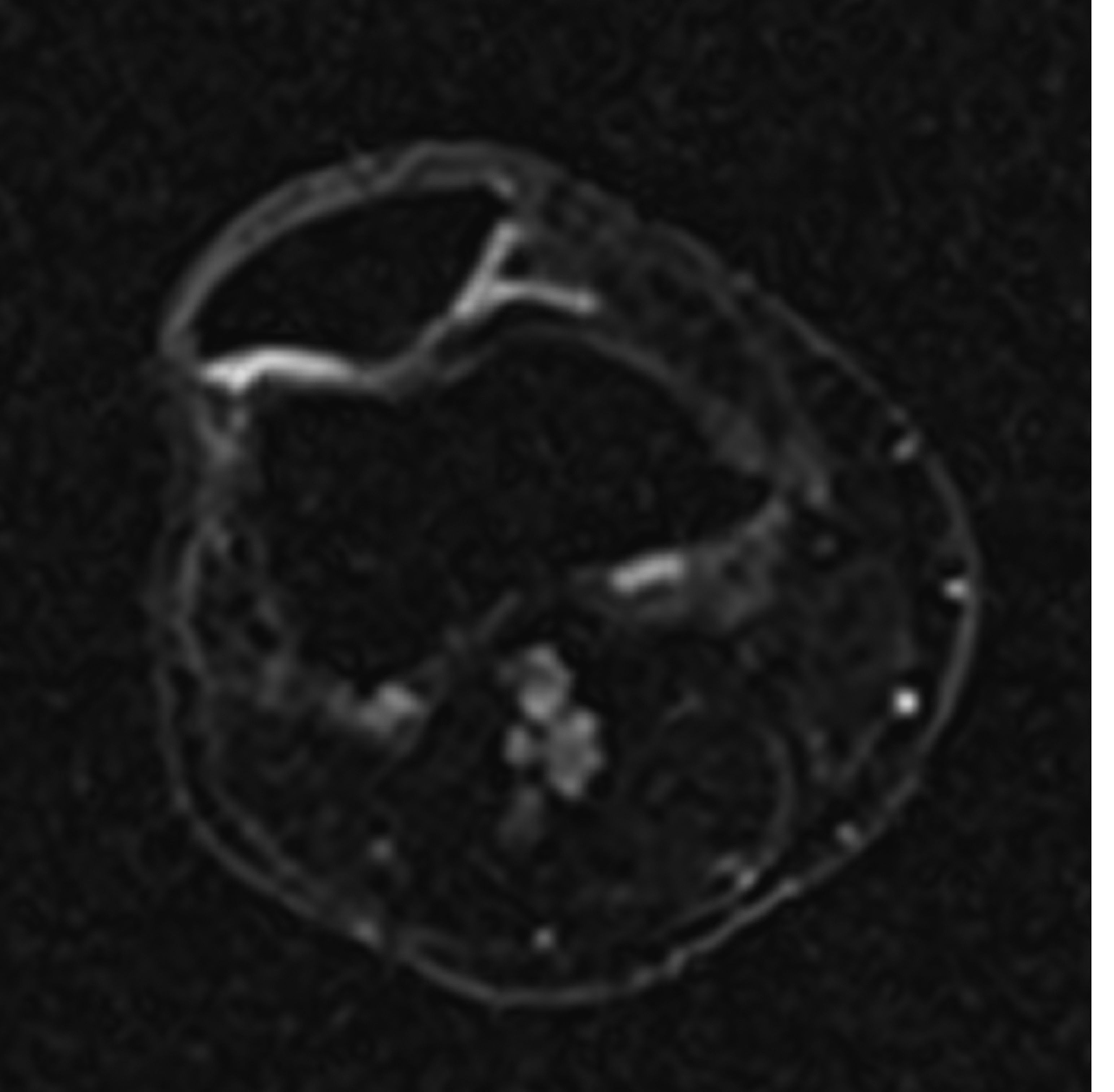
Sodium image in the axial plane of the patella shows the patellar cartilage. At the border from the medial to the lateral facet of the patella an area with decreased sodium signal-to-noise ratio (SNR) is visible which corresponds to a decreased content of glycosaminoglycan (GAG) although the cartilage thickness is preserved. This means an early stage of cartilage degeneration in this area with a focal loss of GAG.
(Provided by Prof. Siegfried Trattnig and the MR Centre of Excellence)