1. Radiation pneumonitis
2. Tuberculosis
3. Carcinoma of the lung
4. Pulmonary hypertension
Answer to case # 2


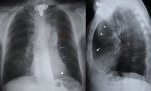
The first impression when looking at the PA chest radiograph is a prominent left hilum (red arrow). If we continue to look (we have learned to fight satisfaction of search/sex, muppet dixit), we discover that the left heart border is indistinct (blue arrows), whereas the aortic knob is well defined (luftsichel sign) and there is tenting of the left hemidiaphragm (white arrow) also called the juxtaphrenic peak sign.
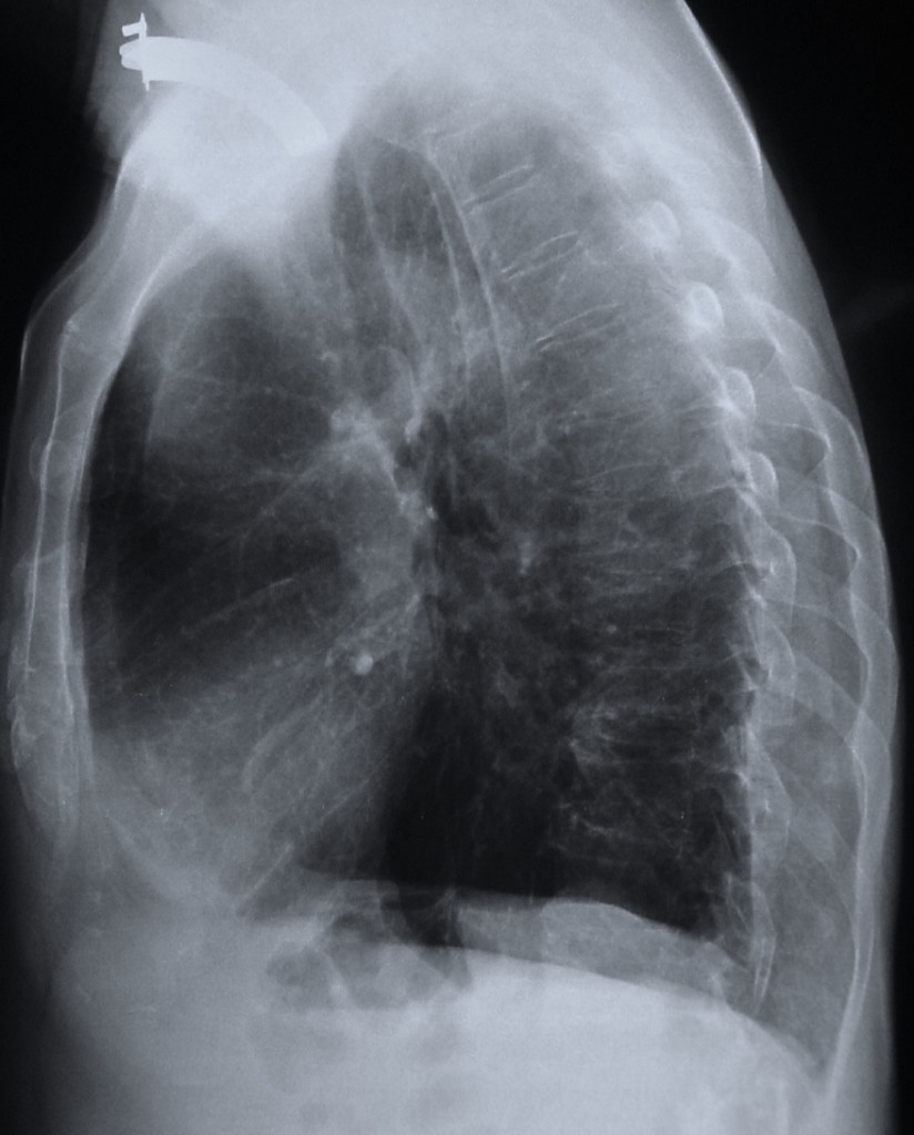
All these are typical signs of left upper lobe collapse. Diagnosis is confirmed on the lateral view, which shows a thin line behind the sternum (arrows), representing the left major fissure displaced anteriorly due to the marked loss of volume of the left upper lobe.
Once the collapse is recognised, carcinoma of the lung is by far the most likely diagnosis, since the majority of collapses of the left upper lobe are due to carcinomas.
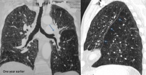
An interesting fact is that this patient had a CT one year earlier, read as normal. Review of the CT shows the endobronchial tumour (arrow) and moderate loss of volume of left upper lobe in the sagital reconstruction (arrows), both overlooked at that time.
Teaching point: important to know (and recognise, says the muppet) the signs of left upper lobe collapse because the great majority of them are due to endobronchial carcinoma
 Clinical history: asymptomatic 75 year-old man, operated on ten years ago for carcinoma of the larynx. Current chest radiographs were obtained during the yearly routine control.


radiation pneumonitis
Tuberculosis
Carcinoma of the lung
Radiation pneumonitis
Tbc
Carcinoma of the lung
I think it looks like post radiation therapy
pulmonary hypertension
Radiation pneumonitis & Carcinoma of the lung.
On PA chest: opacity- post rib6 right,and between 8-9 left.
pulmonary hypertension
Radiation pneumonitis?
left sided hyperlucency with peribronchial thickening in the lower lobe. prominent left hilum -?mass/lymph node.
old fibrotic changes right apex.
pulmonary hypertension
the given case of the gentle man probably indicates a tuberculosis feature based on a dense oval lesion on rt upper lobe and the lateral view indicates signs of pneumonitis and sclerotic changes on base and lower lobe of lungs
final diagnosis : tuberculosis ,radiation pneumonitis
the case is about a 75 year old man who has been operated 10 years ago for carcinoma larynx. he is asymptomatic and for routine chest xray done
i think the most appropriate answer is radiation pneumonitis as changes in his lungs are generally confined to the field of irradiation.
observe that there is a diffuse haze in the irradiated region with obscuring of vascular outlines.Patchy consolidations appear in RUZ and then coalesce to form a relatively sharp edge that conforms to the treatment portals rather than to anatomic boundaries.
fibrous change in RLZ and overlying left hemidiphragm seen as tenting
small amount of effusion is seen as there is blunting of anterior CP angles in lat chest view which could be reactionary to the radiation changes in this patient
active infection is not seen in this case and ca lung doesnt fit in as changes are not there
no e/o pulmonary hypertension
i put my money on radiation pneumonitis
kindly u throw light and inform us abt the right choice and reason it out please
radiation pneumonitis
Dear Dr. Sharma, what do you think of the retro-sternal line on the lateral chest?
Pepe! How fun!
collapsed left upper lobe
Radiation pneumonitis
I’m stuck in these details: translucent contour of lower left arch of heart, vertical opaque band trough the shadow of heart+mediastinum – left contour better defined (no correspondence in lateral seen by my eyes 🙂 ).
What I see: thickening of pleura in left costophrenic angle (anterior mostly), ill-defined contour of upper mediastinum+lost of translucency in upper pre-mediastinum compartment – lateral view (=radiation pneumonitis following larynx Ca. treatment??), ovoid opacity in left hilum well defined only on left side (AP view)=belonging to mediastinum, calcified round lesions (middle area of right lung and right hilum) (=scars of TB?), tracheostomy (following larynx Ca.), osteoporosis (vertebrae mostly).
If seems like a too long comm, I apologize but I need (expect from readers) clear directions for my “radiological” thinking.
Thanks in advance! AE
Dear Wisper in the wind, I think you describe very well the findings. All you have to do now is to put two and two together.
Sorry, I cannot keep on because I do not want to give away the diagnosis.
Keep up the good work
Tuberculosis!
radiation pneumonitis
yeah,agree with the whisper in the wind…stigmata TBC,but that sharp and continuity contour of the left hemidiaphragm,and paravertebral sharp line maybe suspicious from litlle bit of pneumomediastinum(tracheostomy),or postradiation fibrosis…scolyosis of the vertebra too.
right heart ventricle dominated,because of COPD…no Neo signs 4 me
Are you sure?
well,translucency on the left was due to colaps of the left upper lobe,but i’m proud of seeing that signs…TBC lymph nodes was detail which made a dilema,and that sign of major incisura,i think it was a pleural athesia 🙂
anyway,thank you mr. profesor for nice presentation and replying comment
Dear friends, i am very pleased with your answers. Some are right and others are not.
Perhaps is too early to post the final diagnosis. Do not want to spoil the chances of the late comers.
Anyhow, i would exclude the diagnosis of pulmonary hypertension. Basic findings in PH are prominent hila, enlarged main pulmonary arteries and cut off of peripheral arteries. In the present case only the left hilum is prominent and there is a pulmonary infiltrate. These findings make PH unlikely.
So, now you have three choices.
Final diagnosis by the end of the week
Tuberculosis.
Pleural calcification, tenting of left dome of diaphragm with fibrotic band in left lower zone, calcific opacity in right middle zone, ?left hilar lymph nodes.
But in a 70 year old asymptomatic patient, having a left hilar mass..i think he would need a CT to rule out a central tumour.
Radiation pneumonitis
Thank you Professor, simultaneously challenging and educative.
A Really Good Case !!!
Radiation pneumonitis + TBC
Luftsichal sign and juxtaphrenic peak sign & left hilar mass suggest left upper lobe collapse following lung carcinoma