Below are images of a 35-year-old woman with chest pain and progressive dyspnoea for the last three weeks. Leave your thoughts in the comments section and come back on Friday for the answer.
1. Lymphoma
2. Pleural metastases
3. Mesothelioma
4. Any of the above
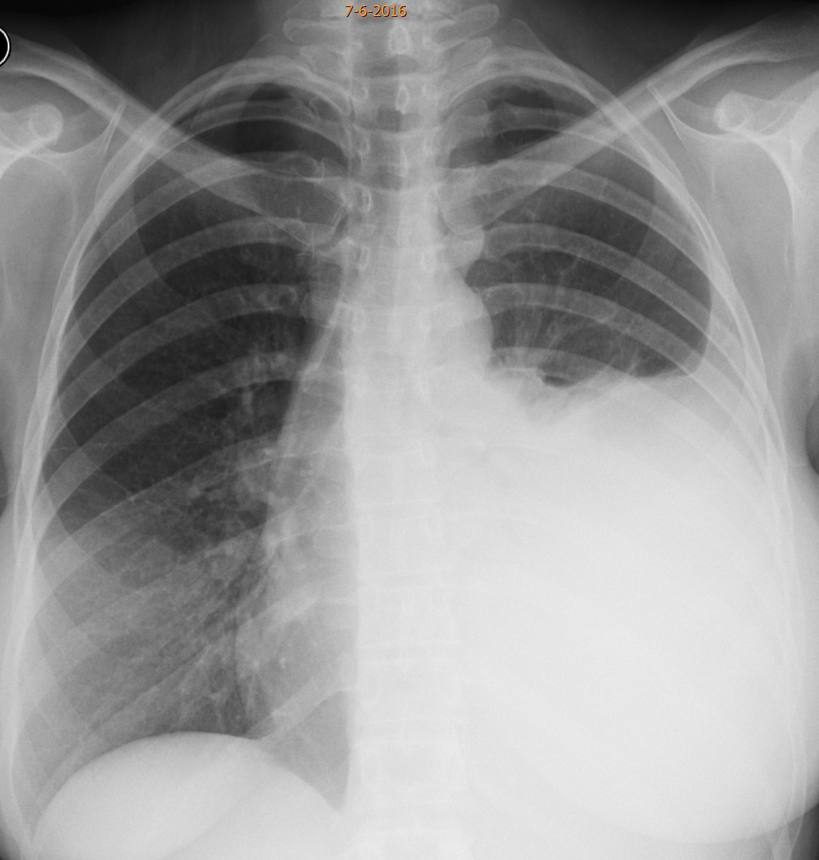
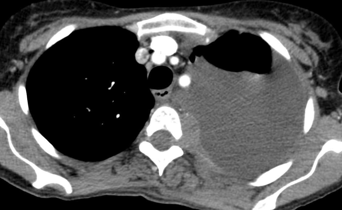
Findings: the PA radiograph shows a large left pleural effusion. A small mass visible in the left upper mediastinum (A, arrow).

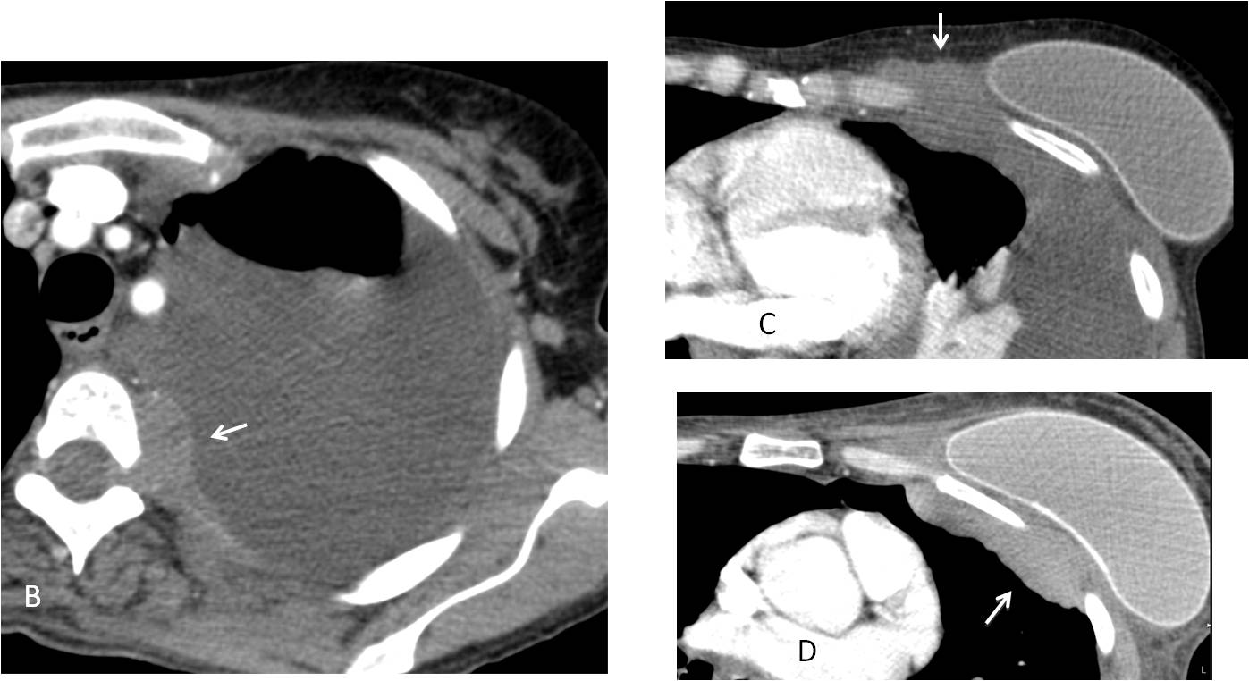
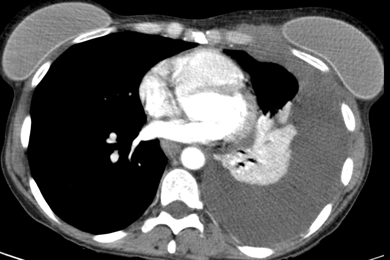
Enhanced axial CT confirms the upper mass (B, arrow), which is invading the neural canal. There is a second mass involving the chest wall (C, arrow), adjacent to the left implant, better seen after the fluid was drained (D, arrow).
I am showing this case because it is a good example of a little known entity: lymphoma developing in women with breast implants. The relationship is not clear, but all reported cases are anaplastic large cell lymphomas, ALK negative.
Our case had these characteristics. The patient had recurrent seromas in the left breast. Cytological examination of the fluid was negative until the patient developed the actual tumour.
The chest radiograph (E) became normal after treatment.
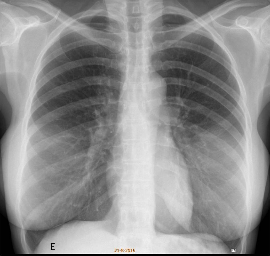
Final diagnosis: anaplastic large cell lymphoma in a patient with breast implants.
Congratulations to Mauro who made a good differential diagnosis, with Mahmoud as a worthy second.
Teaching point: it is wonderful to learn new things! I was shown this case recently by a junior staff and, of course, I had no idea it existed. Whether or not the relationship between implants and lymphoma is real, I am going to look at it from now on. I remember publishing a paper about two patients with implants and recurrent breast seromas and I am planning to review the clinical histories to see what happened.


Hello.
There are no calcified pleural plaques, so I guess the diagnosis of mesothelioma, which is generally related to asbestosis, is unlikely, thus excluding 3 and 4.
I think it could be a metastasis, maybe from breast cancer, as the patient has bilateral implants (reconstructive?). Plus there is some soft tissue medial to the left implant which is not present on the other side. And there is an axillary lymph node on the left which doesn’t`t look very typical.
Nevertheless the appearance of the lesion makes me think of lymphoma, as it is kind of plastic and protrudes to the neural foramen.
So I`ll go with lymphoma.
In the PA Rx view there is a moderate inferior pleural effusion, with a well delineated convex superior border (Golden´s sign). The main left bronchious is retracted probably because of parcial LIL collapse.
There is an increased density proyected over both hemithorax becasue of breast prothesis.
In the CT we see a loculated moderate left pleural effusion with an extrapleural posterior paravertebral lesion that is affecting the conjuntion hole (we can see in the PA Rx view). I dont know if we have an increased density posterior to the left breast prothesis.
Why does the patient has breast prothesis? Is only an esthetic desicion or a medical problem?
Pleural metastasis would be a good option.
Breast prosthesis were for cosmetic reasons.
My diagnosis is lymphoma
-Extension into left neural foramen without bony destruction or remodeling is suggestive of soft tumor like lymphoma rather than metastasis
-The bilateral breast implant may be due previous bilateral breast lymphoma, and the soft tissue adjacent to left breast implant may represent residual or recurrent lymphoma.
Breast implants were placed in 2005 for cosmetic reasons.
Hello,
there is large amount of fluid in left hemithorax with atelectasis probably from fluid compression. Becouse of IDoR’s main topic and increased density near medial edge of left breast implant which going posteriorly to chest I suspect connection of these two main findings: fluid and implant. I would go for inlammatory changes from left breast implant.
I can see you are adding two plus two 😉
In the PA view it seems to be an asymetric with an increased volume and density of the left breast. Perhaps an inflamatory condition like empiema neccesitans would be a good option, and the posterior mediastinal lesion is an incidentally finding (probably nervous tumor like schwanoma).
Deos the patient present infection parameters?
No obvious infection
DM perhaps
There is lytic lesion in medial end of right clavicle seen on CXR
this finding favors metastases over lymphoma
The left clavicle was normal in other radiographs. No lytic lesion. Sorry.
….professore insigne…..linfoma pleurico senza alcuna immagine adenopatica a livello ilo-mediastini o…..non ci credo……mesotelioma pleurico in un soggetto femminile di 35 anni….non ci credo…..il versamento pleurico allora è’ di natura neoplastica per infiltrazione della pleura, da cr mammario sx( al lato mediale della protesi) ove c’è’ una massa che annulla il grasso sottocutaneo……Bari un disastro…abbiamo già’ cambiato l’allenatore ! Un forte saluto da Bari…..
Hello!
I think the answer could be pleural metastases for breast cancer.
On PA – left side hydrothorax, confirmed on CT.
On the first CT image there is mass in the anterior chest wall which grow both inside and outside of chest wall, outside it is close to breast implant.
On the second CT image there is slightly contrast enhancing mass near the body of thoracic vertebrae – it seems this mass is growing in vertebral canal through the foramen intervertebrale.
It seems that it could be pleural mts in anterior chest wall according to the previous breast cancer with the combination with mesotelioma of the posterior chest wall
The case of two histologically different malignancies
Do not make life difficult. Use the KISS method 😉
Any of these
Pleural effusion of left hemithoraks with compression of lung parenhyma next to it.
Fluid in the medial aspect of the left implant with thickening of subcutaneous tissue with fat stranding.
These two findings seem to communicate through the anterior thoracic wall- chronic imflammatory response to implant??
I am not sure if there is thickening and calcification of the pleura in the posterior and medial aspect of the left lung? Or an mass seems to go through the left neural foramen??