Today we are presenting a pre-op chest radiograph of a 70-year-old man with carcinoma of the bladder.
What do you see?
Check the image below, leave your thoughts in the comments section and come back on Friday for the answer.
The preoperative chest radiograph shows left diaphragmatic and pleural calcifications (A, arrows). There are several opacities in the left hemithorax that may be pulmonary infiltrates (A, red arrows).
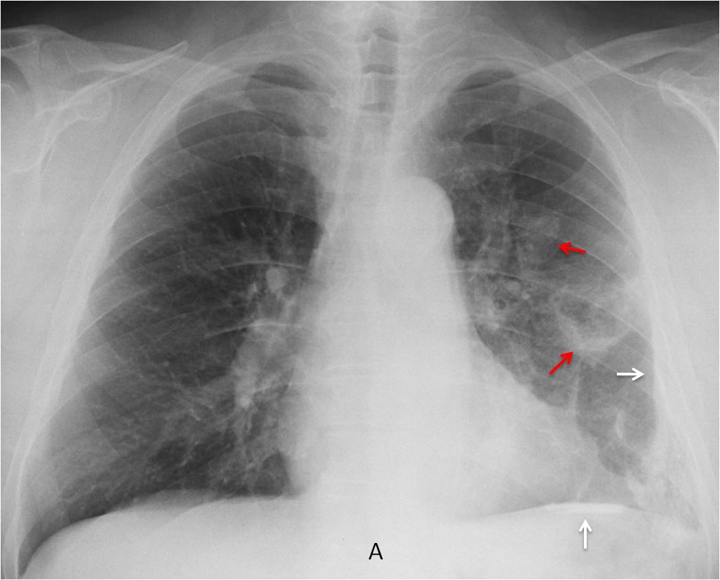
Two considerations should me made: 1) Asbestos exposure is very unlikely because the pleural calcifications are unilateral. 2) The apparent pulmonary infiltrates could be due to pleural calcifications depicted en face.
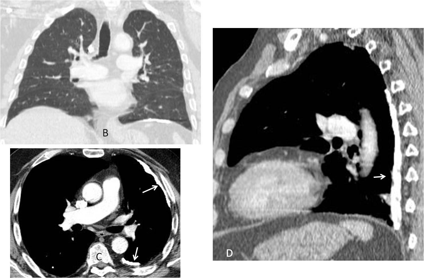
Coronal CT (B) does not show any lung abnormalities. Axial and sagittal enhanced CT shows extensive pleural calcification (C-D, arrows).
The patient had antecedents of TB in his youth.
Final diagnosis: Calcified pleura secondary to previous TB
Congratulations to Nacer, who was the first to suggest the diagnosis
Teaching point: Remember that diseases of pleura/chest wall may simulate pulmonary lesions in the chest radiograph


There is a loss of volume of the left hemithorax.
I see a cavitate lesion in the LLL and an increased diffuse density in this hemithorax, with an obliterated costophrenic sinus. Also, there is a left pleural thickening and a partially calcified left diaphragm.
I think that there is a flattened of both hemidiaphragm.
Granulomatous or occupational dissease would be a good option.
Any interesting personal medical history?
Not really 😉
Cavitating masses in left lower lobe with minimal left pleural effusion. Prominent vascular markings in left upper lobe and peri hilar region with .? Bulla in left lower lobe
Asbestosis?
Same like MK and calcarea pleurae because of status post Tbc pleuritis.
….professore stimatissimo……l’anamnesi lavorativa potrebbe essere la chiave interpretativa tra le lesioni “pleuriche” nell’emitoace S ed il cancro della vescica….il cancro vescicale è’ secondo per incidenza tra i tumori professionali dopo quelli per esposizione ad. Amianto…..l’esposizione ad amine aromatiche, prodotti in vari tipi occupazionali, può avere determinato gli esiti pleurici ed il successivo cancro della vescica….il Bari ha finalmente vinto una partita….per il Barca senza Messi è’ dura…..per ora il Real è’ in testa….
I am doubting about cavitary lesion va pleural plaques that are more typical in abestosis
cavity left mid zone.
pleural thickening + suspected effusion left CP angle and base.
foci of pleural calcification including above dome of diaphragm.
cannot exclude rib lesion.
any history of asbestos exposure, please?
No asbestos exposure, but you should have guessed 😉
Perhaps, hystory of “talcodesis”?
No talc placement
Thick calcified left pleual plaque + cavitary lesion in in left lung with left pleural thickening at left costriohrenic angle + no history of asbestos exposure—–raise possibility of TB.
Sequellar lesions of left pulmonary and pleural tuberculosis
bladder cancer with calcified pleural mets on the left (chondrosarcomatous component). Rare but recognized.
I googled bladder cancer and calcified pleura!
Never saw a case, but I guess it’s possible. Goes against the KISS method, though
Just the chest x ray – 1st ddx would be granulomatous dis like TB.
Trying to correlate with bladder ca – the only thing i can think of schistosomiasis
one other possible link – bladder cancer treated with BCG gets TB from the cancer treatment due to being immunocompromised and then has pleural calcification secondary to the TB.
Ingenous but unreal 😉
Sorry what is the KISS method?
KISS is an acronym for “Keep it simple, stupid”. It emphasizes that simple explanations are better to interpret the findings than convoluted ones. It is similar to “Occam’s razor”: when there are two explanations for an occurrence, the simple one is the best.
thank you