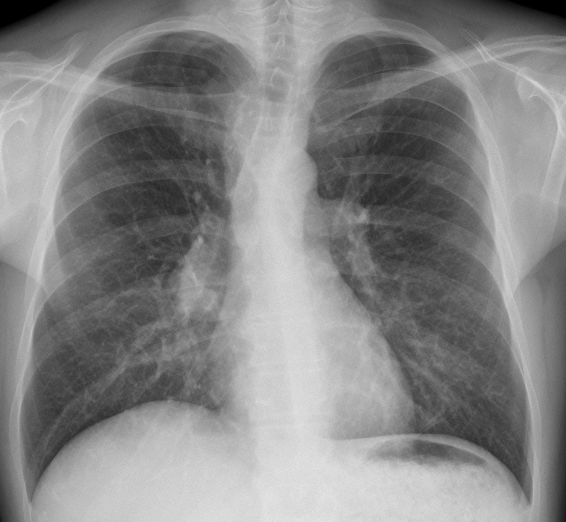
Today I am showing a PA radiograph of a 48-year-old smoker with a persistent cough. What do you see?
As in last week’s case, I will show additional images on Wednesday morning.
Check the images below, leave your thoughts in the comments section and come back on Friday for the answer.
As promised, here are the two additional images. Do they help you to reach a conclusion?
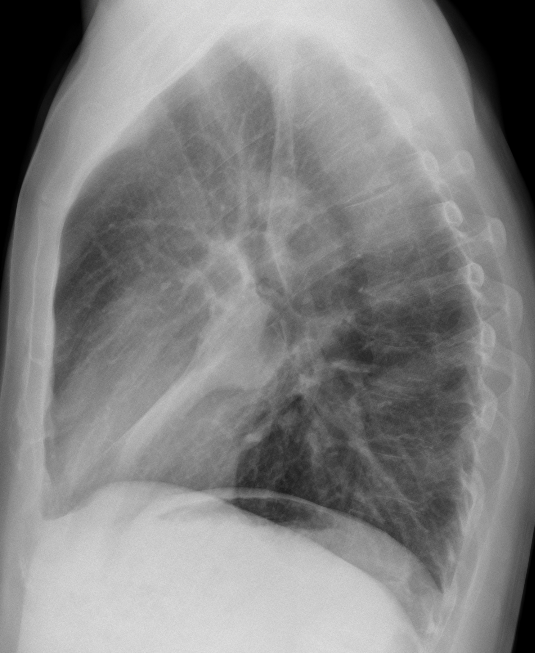
Findings: PA radiograph shows a well-defined nodular shadow superimposed to the right hilum (A, arrows). Lateral view shows a rounded mass in the right hilum (B, arrow), causing marked collapse of the RML (B, red arrow).

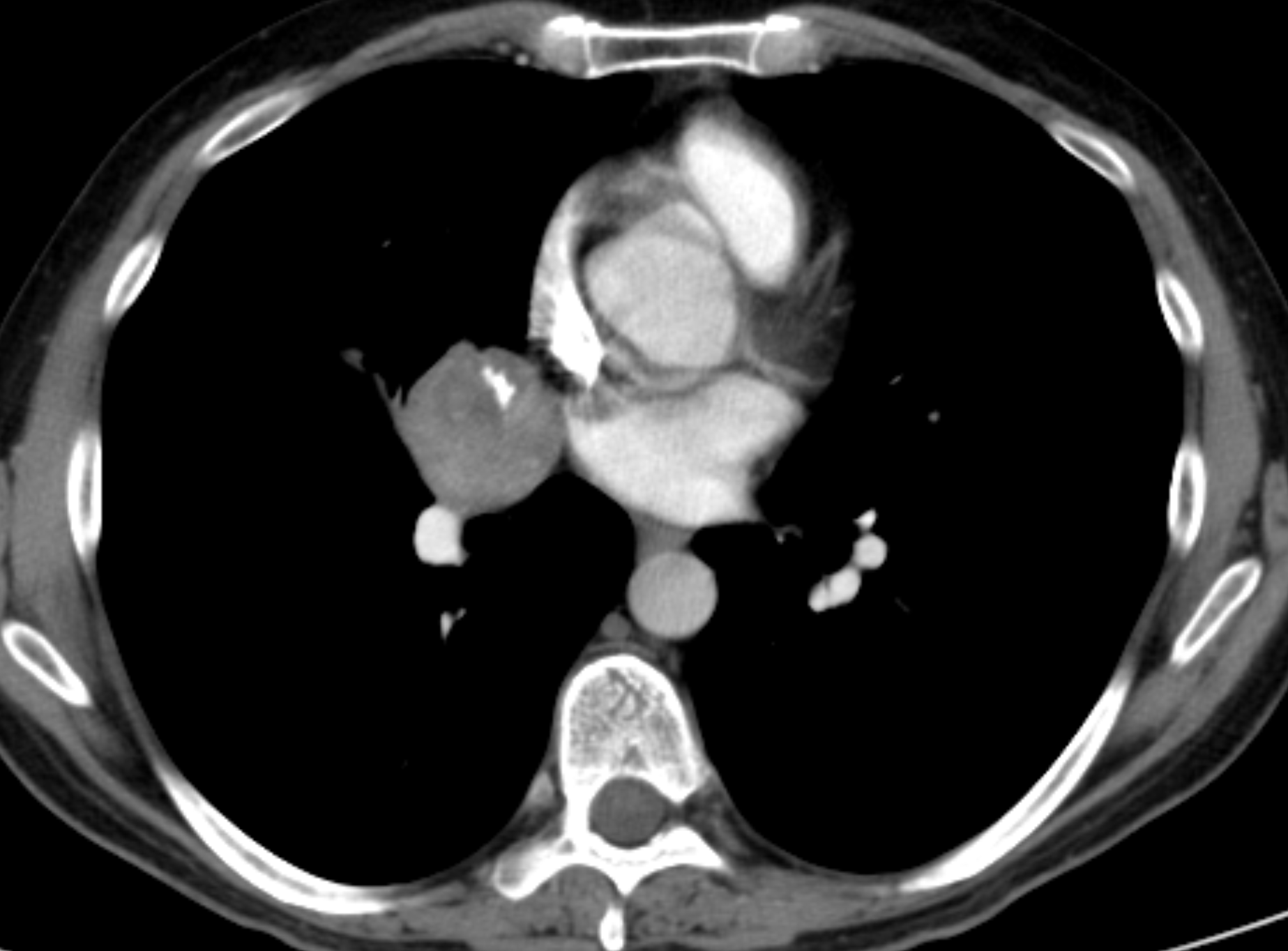
Sagittal CT confirms the mass (C, arrow) and the RML collapse (C, red arrow).
There is coarse calcification within the mass (C-D, yellow arrows). PET-CT shows marked uptake of the mass (E, arrow).
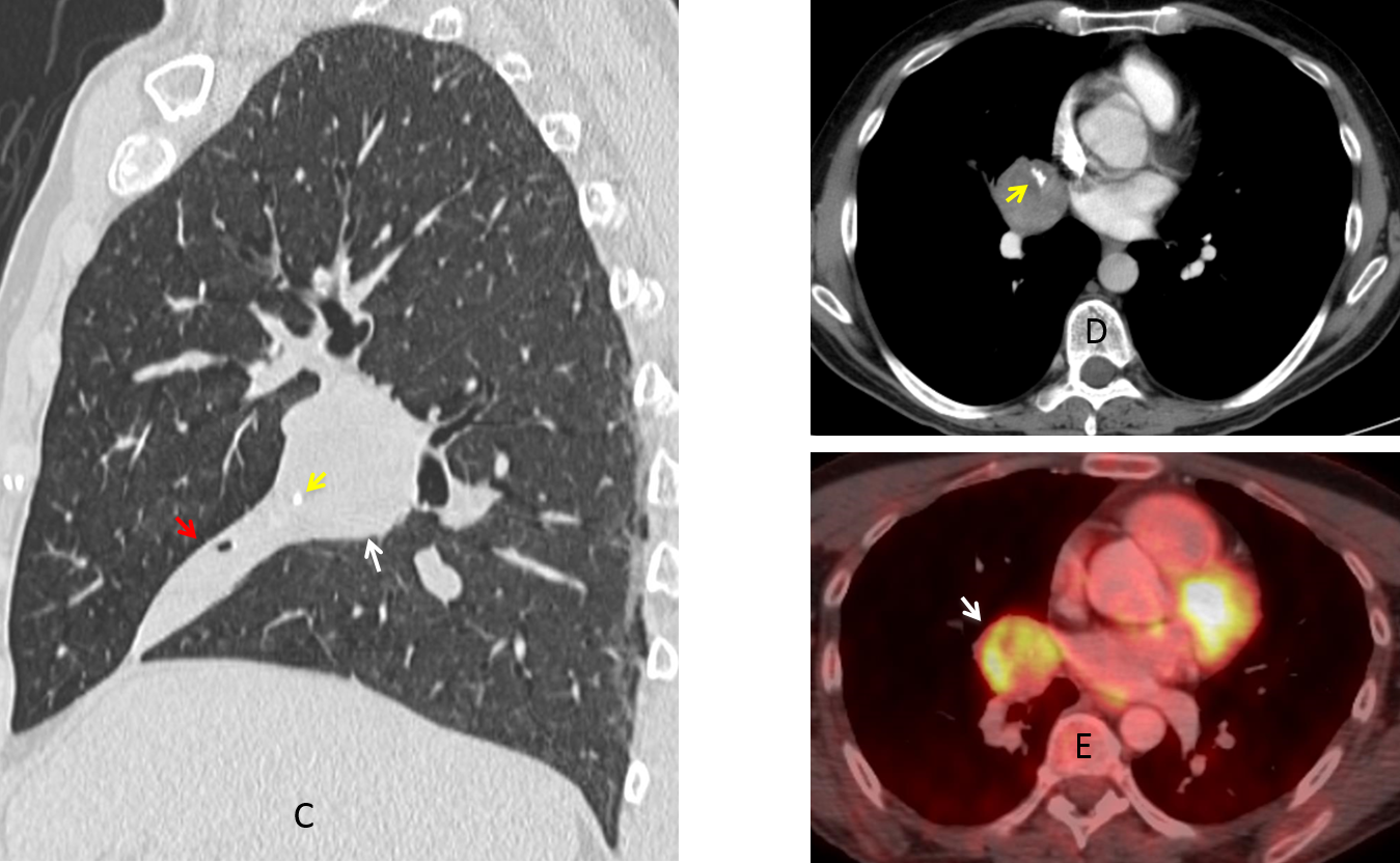
Bronchoscopy confirmed a mass occluding the RML bronchus. Biopsy returned the diagnosis of carcinoid.
Final diagnosis: carcinoid tumor causing marked collapse of RML.
Congratulations to MK, who was the first to mention the correct diagnosis.
Teaching point: this case confirms the importance of the lateral view to complement the information obtained in the PA radiograph. And remember that a nodule with coarse calcification in CT practically excludes bronchogenic carcinoma.


Good morning!
Prominent hilum. There is a subtle hign density proyected over the right hilum with a well defined inferior and lateral borders. Not sillouette sign. Thickened of the paratracheal line and right inferior tracheal desplacement.
Intrapulmomary parahiliar lesion?
Another option will be bilateral hiliar and paratracheal adenopathies like in sarcoidosis
Look at the new images
Round hiliar lesion with calcium that causes segmental ML atelectasi. Carcinoid lesion?
I cannot find remarkable brochial thicking and abnormal shadow suspect pneumonea, bleeding, toumour and so on in lung field.
no pleural effusion.
no abnormal tumour in chest subcutaneous tissue.
Only susupect bilateral hilar lymphadenopathy, especially right side.
(high density mass suspected but difficult judgement)
So usual doctor need Chest CT and other factors check.
48-old smorker may have many problem in his life.
Welcome! What do you think about the new images?
Too forward maybe… let’s try it anyway: right hilar/mediastinal bronchogenic cyst
greetings,
– Large faint opacity with round lateral margin over the right descending pulmonary artery, anterior or posterior to it.
– Thickening of the right para tracheal stripe.
DDX: neoplasm with lymph node involvement, infected cyst with reactive lymph nodes.
There is a round right hilar mass. The right heart border appears sharp along with positive hilum overlay sign indicating the mass is most likely mediastinal in origin, probably anterior. I would be thinking of thymic/bronchogenic cyst vs lymphoma/thymoma. CT will be helpful for differentiation.
There is an opacity with well-defined outer and lower margins and not well seen upper border. I agree with others, and would like to add the mass of chest wall; neurinoma
It is a sharply demarkated mass overlapping on the right hillum, because hillar structures ( vessels , bronchi ) are well visible so it means that this mass must be in front or behind the hillum. Main pulmonary vessels are enlarged . There is also an opacification in the middline just over the carina, quite round and well demarcated.
For better answer left lateral view recomended.
Now you have a lateral view and a CT. What do you think?
Chest X-ray frontal projection of an adult patient shows a faint opacity in the right hilar region,it shows well defined inferior borders, it does not obscure the hilar vessels nor the right heart border and shows acute angles with the mediastinum. There is no calcification or cavitation, the adjacent bone structures are normal. The opacity is mostly intrapulmonary, differential diagnosis is wide, I would like to review the lateral chest X-ray and compare with old films.
(I’m not good at English)
PS
After reading other comments, I suspect right side mass more than BHL.
Like this case, if permitted, Very low dose CT is suitable than usual CT.
Because the points of differencial diagnosis are…
Bronchial problem or not
inter-mediastinam or not
BHL or not
Greetings,
In the lateral view the mass is causing segmental collapse, probably in the anterior lower lobe.
In the CT image the mass looks mildly heterogeneous with probable fat density within it, and an irregular eccentric calcification.
Impression: the lesion is probably a hamartoma causing a segmental lower lobe collapse, but I think bronchogenic carcinoma should be included in the differential.
Dont you find it strange that a central mass is causing a segmental collapse?
HU measurements did not find fat.
Greeting,
I can see a small tail emerging form the lateral side of the lesion with subtle heterogeneous enhancement and central hypodensity adjacent to the dense area/calcification.
I would consider a thrombosed aneurysm in a segmental artery with a residual lumen that is avidly enhancing and looking like a calcification.
Another option is the bronchial carcinoid with eccentric calcification.
….AMARTOMA ENDOBRONCHIALE CON ATELETTASIA LOBO MEDIO…Saluti dalla PUGLIA…
I agree with genchi bari italia – calcified endobronchial hamartoma, indolent lesion
The tomour has calcified lesion and clear border with surroundings.
So, I suspect hamartoma or teratoma or other benign tomours.
But I have some difficulty about diagnosis because of continuous lesion,
maybe thickness of lobes or some fluid, and toumour is enhanced not equally ( looks like tomour in tomour).
So I need other figures and informaton.
Your analysis is correct. Final diagnosis was carcinoid tumor.
Thank you, Sir.
A general question… Why a left lateral is taken in this case? And if no side is specified which projection do you perform?
Thanks
We usually take a left lateral projection because it is our routine. And I am accustomed to look at the radiograph with the spine on my right, so I always place the image in that position.
Thanks for participating 😉
I cannot find remarkable brochial thicking and abnormal shadow suspect pneumonea, bleeding, toumour and so on in lung field.
Greeting,
I can see a small tail emerging form the lateral side of the lesion with subtle heterogeneous enhancement and central hypodensity adjacent to the dense area/calcification.
I would consider a thrombosed aneurysm in a segmental artery with a residual lumen that is avidly enhancing and looking like a calcification.
You can have an astounding and unparalleled experience to unwind and they ensure that each penny you spend doesn’t go waste. Free female Escorts in Delhi are notable for their administration on the off chance that you are in the city interestingly and feeling forlorn while visiting new places at that point book them they will make your visit most noteworthy. Huge quantities of models are full time utilized and some of them are doing part occupations.
Thanks for taking the time to discuss this, I feel strongly about it and love learning more on this topic. If possible, as you gain expertise, would you mind updating your blog with extra information? It is extremely helpful for me.