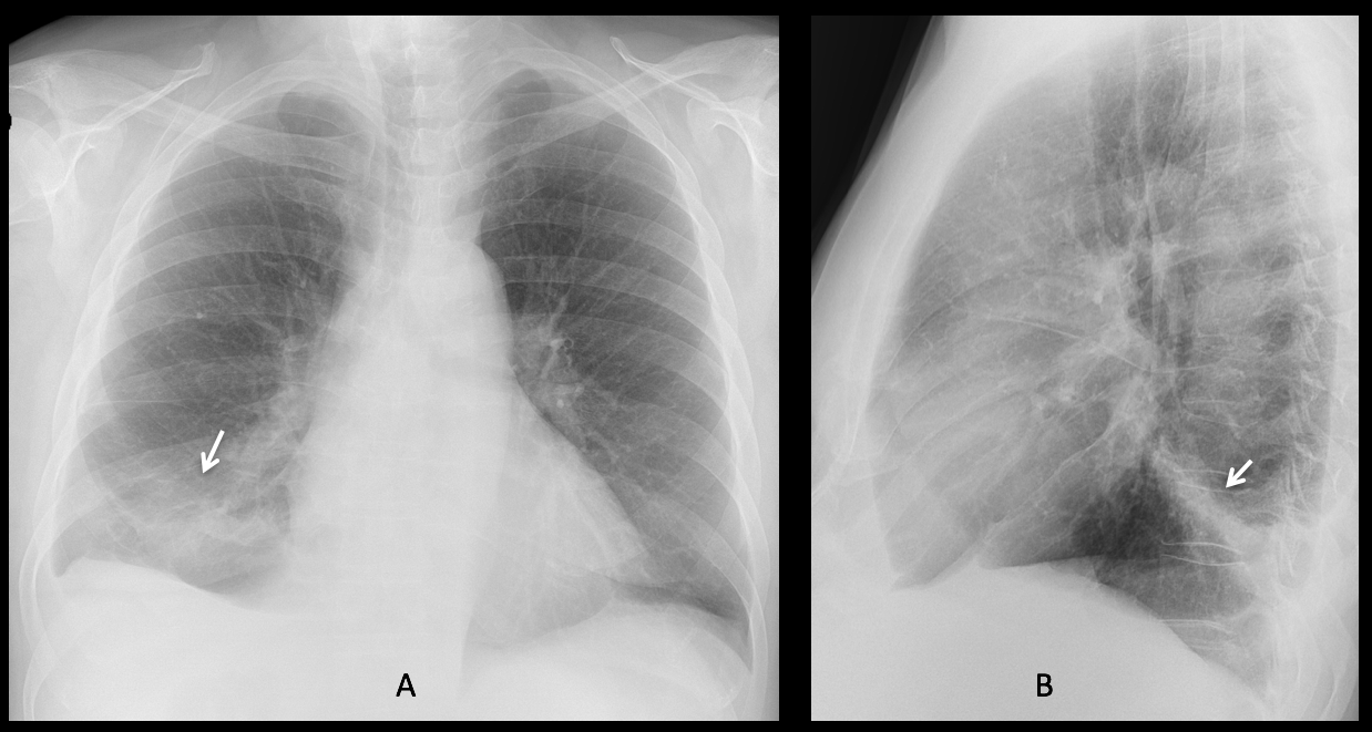
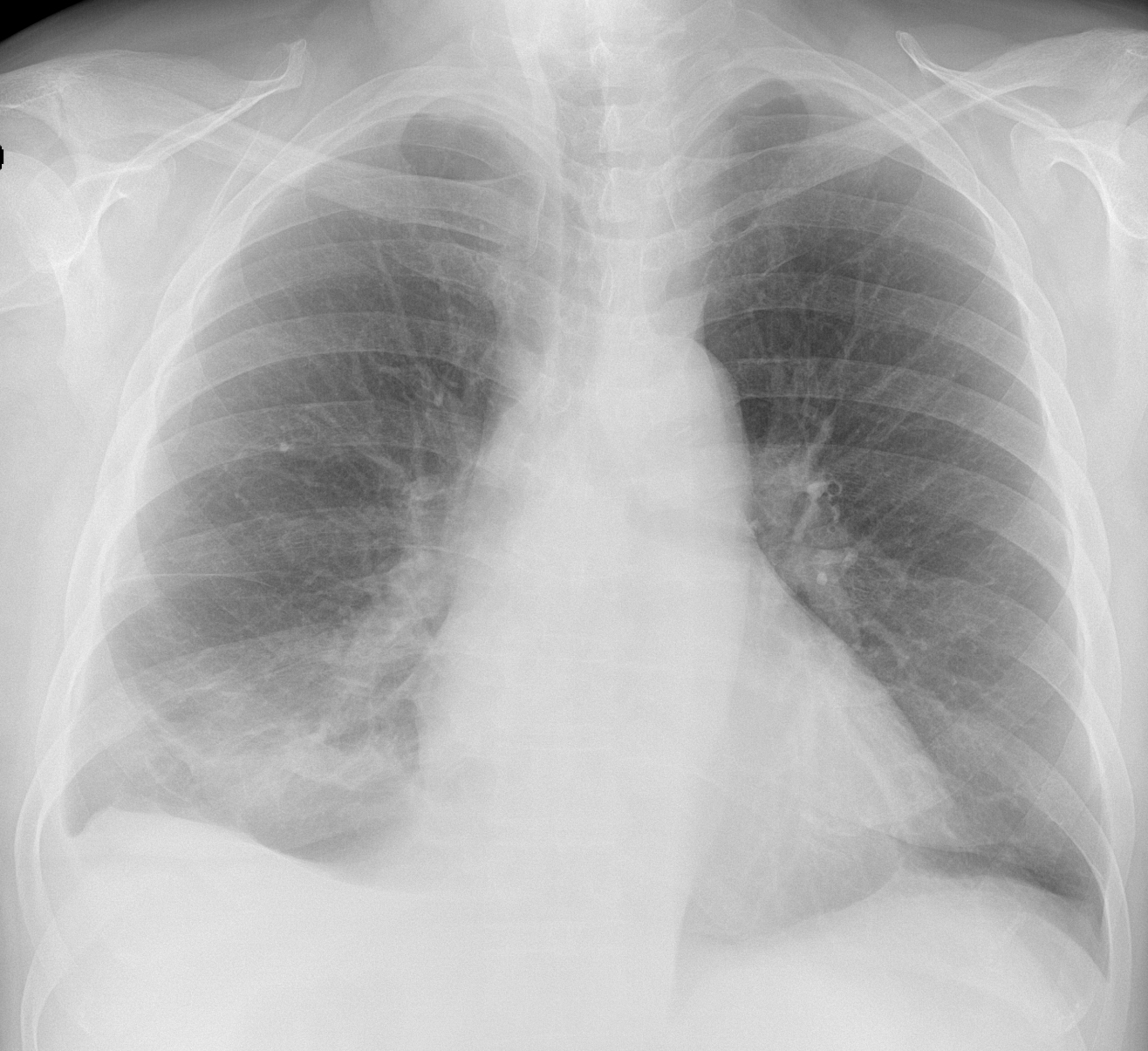
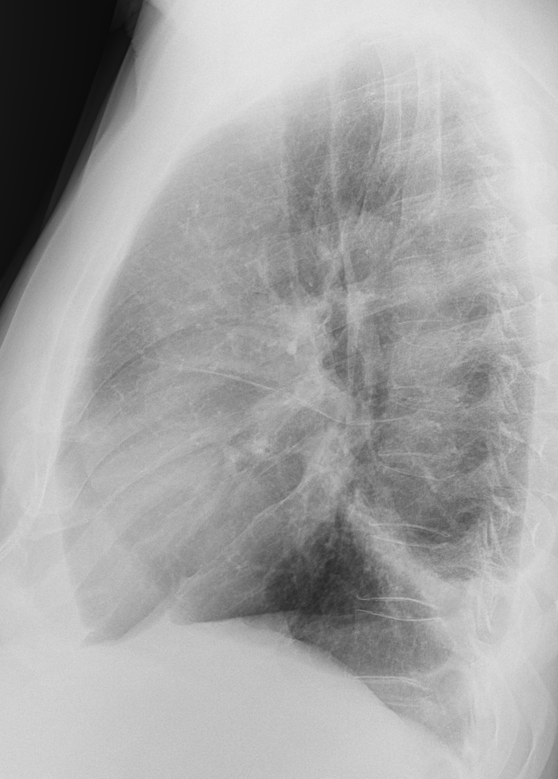
Findings: the PA radiograph shows an ill-defined opacity in the RLL (A, arrow), accompanied by a small pleural effusion. The lateral view shows involvement of the posterior segment of the RLL (B, arrow).
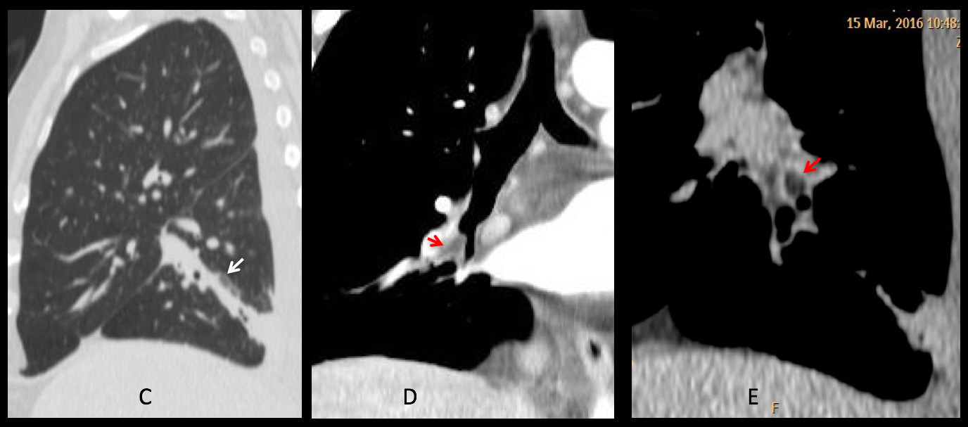
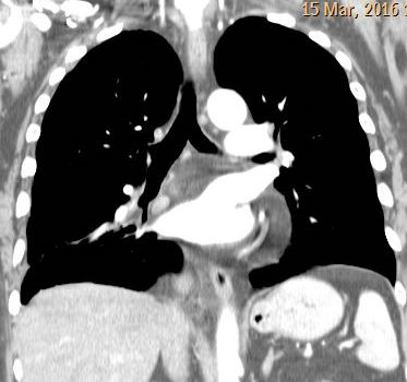
Sagittal CT confirms the segmental involvement (C, arrow). Coronal and sagittal CT with mediastinal window shows a low-density lesion at the origin of the bronchus (D-E, red arrows).
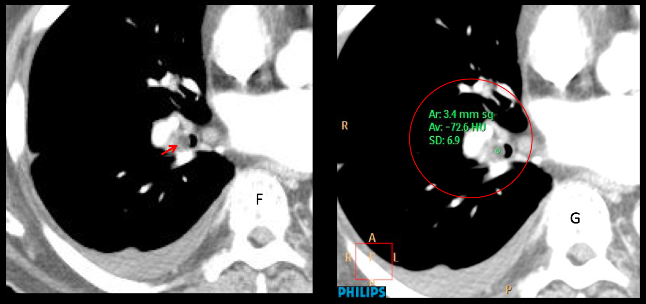
Enhanced axial CT confirms the endobronchial lesion (F, red arrow), which measures -72 H.U. (G, circle). Endobronchial lipoma was suspected and confirmed at bronchoscopy.
Final diagnosis: endobronchial lipoma of posterior segmental RLL bronchus
Congratulations to MK and MP who, with a little bit of help, made the correct diagnosis.
Endobronchial lipomas, also called lipomatous hamartomas are not infrequent in my experience and easily diagnosed by CT. As a bonus point, the last figure shows my first lipoma, seen in the eighties and discovered because of the descended right hilum.
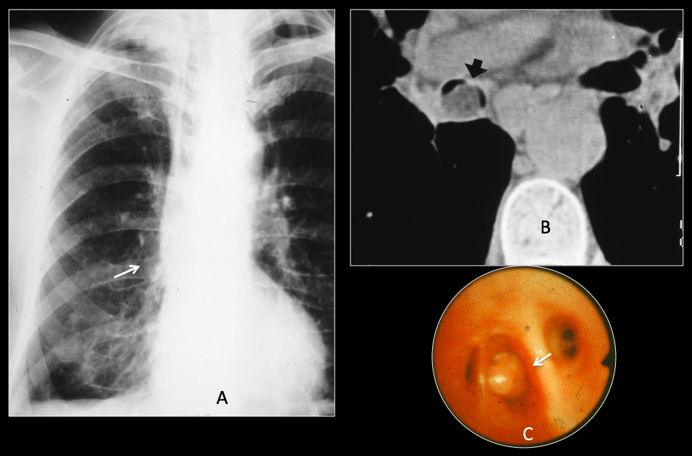
56-year-old male with chronic cough. The right hilum is displaced downward (A, arrow). Unenhanced axial CT shows a low-density mass in the intermediary bronchus (B, arrow). Lipoma confirmed at bronchoscopy (C, arrow).
Teaching point: in lobar or segmental lesions of the lung always consider the possibility of an endobronchial lesion. It led to the diagnosis in this patient who came for an unrelated process.


Good morning!
In the x-rays: Right pleural effussion with an alveolar consolidation with pleural based (infeccion?, pulmonary infarct?).
On coronal CT there is not pleural effusion (different day than x-ray?), but I think there is a pericardiac effusion…
Indeed the attenuation of the pericardial tissues is like fat not water so not pericardial effusion.
In the CT there is an increased attenuation of the right costophrenic fat: fat necrosis? Perhaps the patient has pleural effusion with right segmental atelectasi because of the pain, and now after a few days we can see the fat necrosis.
Has the patient had any thoracic sympton?
You are missing the obvious. There is segmental affectation of RLL. What may cause it?
Endobronchial or hilium process. In the CT the inferior segmental bronchius is ocuppied with a low density, so endoscopic will de made.
And what the endoscopy should show?
Mucus plug?
Or..?
Endobronchial lipoma!
Bronchogenic carcinoma…
Come on, you can do better! How many low-density carcinomas have you seen?
So one possibility would be a bronchial hamartoma which according to the literature is the most common benign endobronchial lesion. CMAJ. 2011 Sep 20; 183(13): E1039
Another possibility is an endobronchial lipoma which according to a literature search would be a rare cause of bronchial occlusion.
….buon Natale…..lipoma endobronchiale con atelettasia ed adenopatie ilo-mediastiniche.
Feliz Navidad!
The frontal radiograph shows inhomogenous haziness of the right lower zone. no silloutment of the cardia or diaphragm. both CP angles are clear, however the right dome of diaphragm is directing laterally (subpulmonic effusion?)
The right lateral radiography is showing linear atelectsis of one of the segment of right lower lobe. No bluting of the posterior costophrenic angle.
coronal CT image is showing a small low density mass in the right lower lobe bronchus.
the diagnosis is endobronchial carcinoid with partial atelectasis posterior segment of right lower lobe.
If I may disagree, it would be very unusual to see a low-density carcinoid, specially after contrast enhancement.