
Dear Friends,
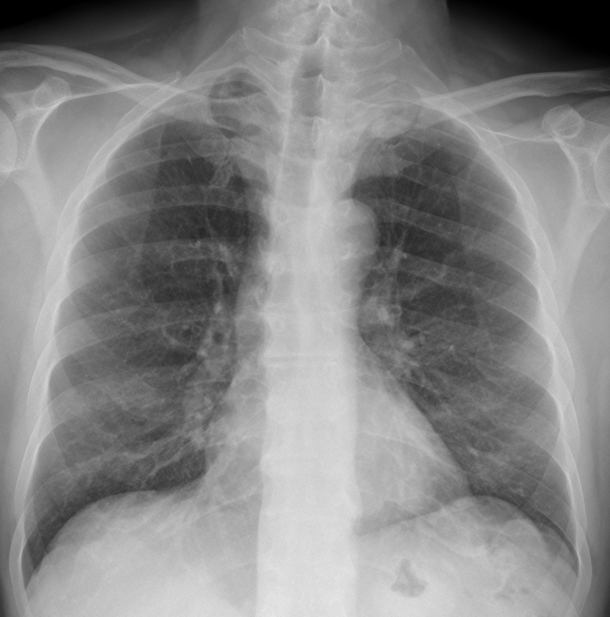
Today, I am showing radiographs of a 68-year-old man with pain in the chest. What do you see?
Check the images below, leave your thoughts in the comments section, and come back on Friday for the answer.
Click here for the answer
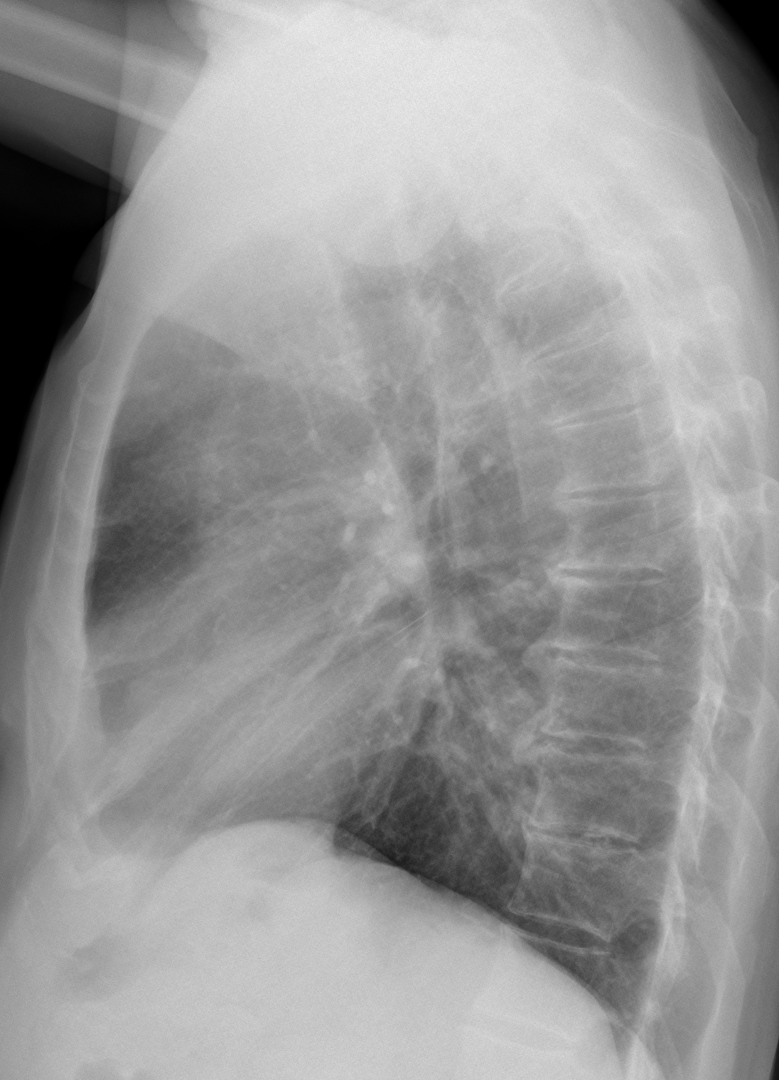
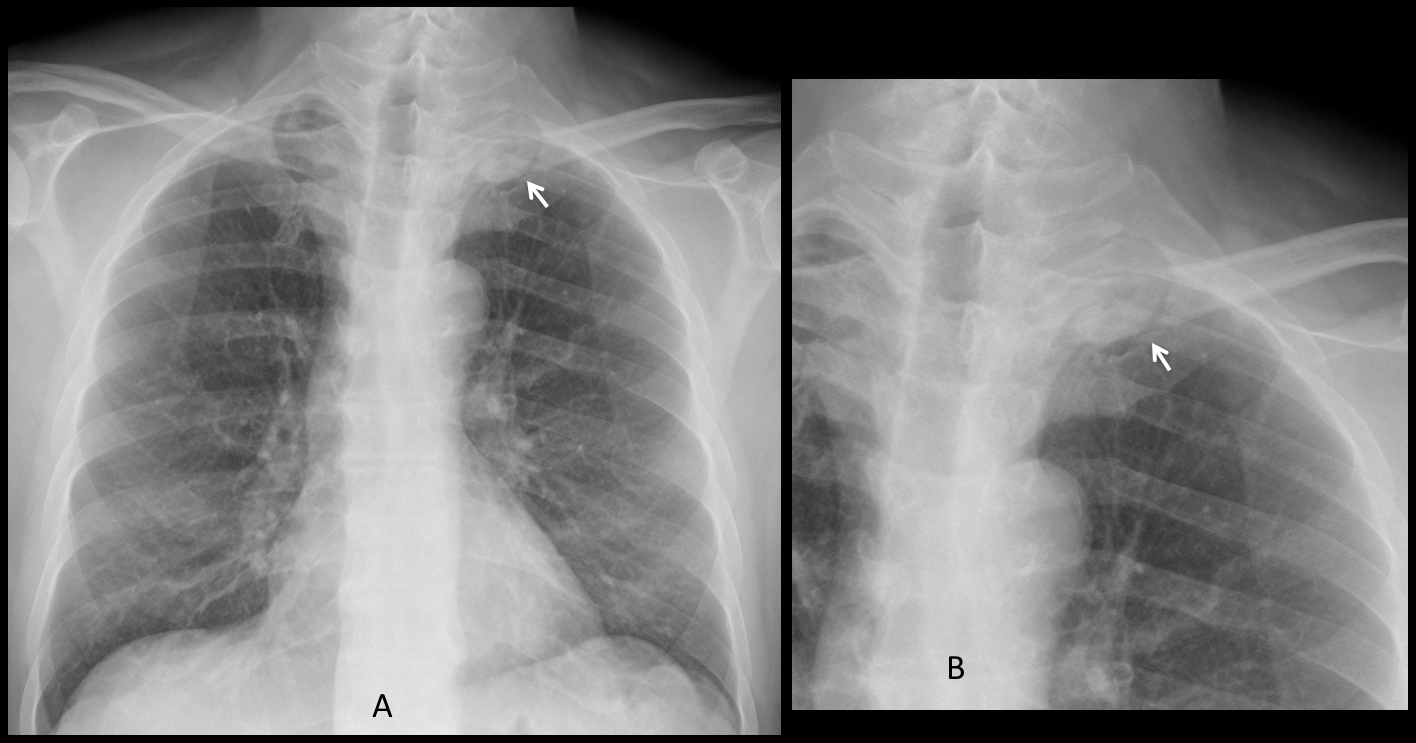
The main finding is a rounded left apical opacity visible only in the PA radiograph (A-B, arrows). The lateral view is unremarkable.
The patient complained of pain in the left shoulder. Coronal and axial enhanced CT conforms the apical mass (C-D, arrows). Needle biopsy came back as adenocarcinoma.
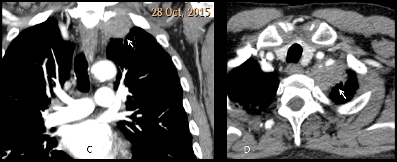
Final diagnosis: Pancoast tumour
Congratulations to the three initial brave correspondents (MK, Coffee and Amago) who made the diagnosis.
Teaching point: the pulmonary apex is a dark area in the chest radiograph and tumours in this location are missed often. Don’t forget to look at the apices in the PA view.


Good morning and happy 2018!
The x-ray is slighly rotated to the left side but there is an increased density in the left apical region (pancoast?). The superior vein cava is more visible than usual because of the slighly rotation.
There is focal mass-like opacity at left apical lung.
And there is suspected increased opacity at LLL zone, retrocardiac region.
Futher CT is helpful.
Left upper lobe atelectasis (triangular homogeneous opacity in the apical region on lateral view) probably due to left sided apical space occupying lesion. Maybe slightly elevated left diaphragm and hilum.
I believe that the triangular opacity is due to the soft tissues of the axilla. If it were real it would be very visible in the PA view.
Vincent, new entry.
Confirm Mk evidence
Increased density in the left apex, if he has pain my first option is a Pancoast.
Not convinced there is malignant pathology at the left apex – it looks bony ? osteophyte.
there is subtle consolidation in the left lower lobe on the lateral best seen on the lateral. This could be the cause of the pain.
Agree with you it looks bony, but very large to be an osteophyte.
The subtle opacity in the lateral view is due to superposition of lower lobe vessels. It is a very difficult area to interpret.