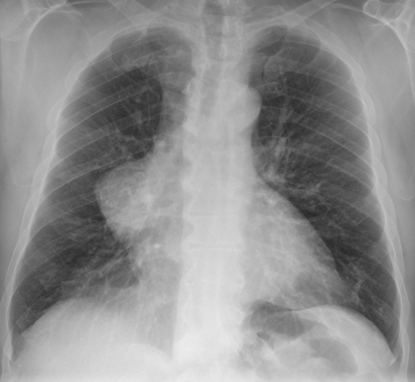
Today’s radiographs belong to a 68-year-old man in whom a chest abnormality was found in a routine check-up.
Check the images below, leave your thoughts in the comments section, and come back for the answer on Friday.
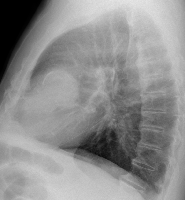
Findings: chest radiographs show an obvious anterior middle mediastinal mass (A-B, arrows) with thick peripheral curvilinear calcification, better seen in the lateral view (B, red arrow).

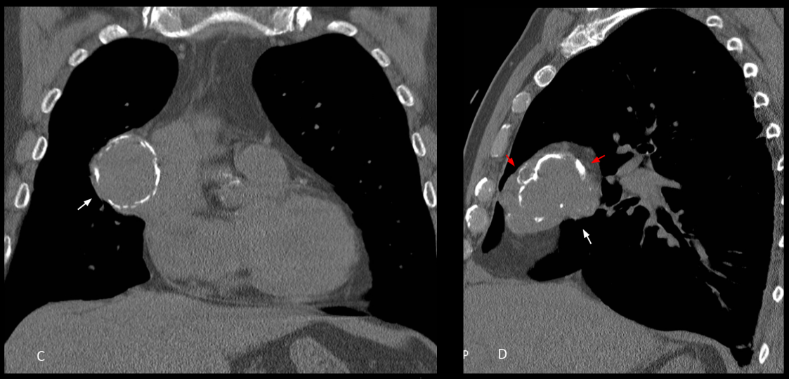
Unenhanced coronal and sagittal CT confirms a solid mediastinal mass with coarse irregular peripheral calcification (C-D, arrows). The irregularity and coarseness of the calcium is better seen in the sagittal reconstruction (D, red arrows).
Diagnosis after surgery was Type I thymoma.
Chest radiographs were not easy to interpret, and I congratulate all of you for trying. In my opinion, the calcium in plain film was too thick to think of a vascular or cystic lesion, leaving open the option for a solid calcified lesion. In my absolutely biased opinion I wish to congratulate Adel as the winner.
Final diagnosis: Calcified thymoma
Teaching point: remember that thymomas may travel downwards and be found in the middle or lower anterior mediastinum.
Also remember that we have eradicated hydatid cyst in Spain!


Hello!!
There is an increased round density next to right mediastinum (sillhoute sign) with calcified margins… pericardical cyst?
I think there is another nodular lesion lateral to the paraspinal line…
I believe the second lesion represents the confluence of right pulmonary veins 🙂
The interrumption of the horizontal fissure at lateral view, makes me think about a pulmonary origin of the oppacity, but… it has obtuse angles with the mediastinum so it should be mediastinal.
There may be some deformation of the cardiopericardical silhouette, w/ calcificacion. ¿Auricular pseudoaneurysm?
Good afternoon sir. The right cardiac border shows increased opacity with convex lateral smooth margins. The lateral view shows a curvilinear calcific opacity on its superior aspect. The RPA is seen coursing through the opacity with no vascular abnormality. On lateral view the opacity is seen to lie in the region of the cardiac chambers.
Likely differential is
1.right atrial aneurysm with calcification.
2.pericardial cyst.
3. Anterior mediastinal mass of cystic nature.
Regards
The appearance of the calcifications suggests the presence of multiple layers in the PA projection. On the lateral projection there is a clear calcific “shell” and smaller rounded opacities anterior to the main one. I think this could be an Hydatid cyst.
Hydatid cysts of lung do not calcify
I agree with you about hydatid cyst of the lung not calcifying. The appearance of the lesion suggests a mediastinal mass and hydatid cyst of mediastinum may calcify
Rt atrial dilatation
Cystic mass showing calcification most probably hydrated cyst
The lateral view the lesion in anterior aspect,with crescent of calcification ..c.t scan examination is needed
But primarily I may say thymoma or pericardial cyst.
Pericardial cyst
Lads calcificaciones perifericas a una lesión de baja densidad son frecuentemente observadas en quistes. Muy probablemente se trata de un quiste pericardico.
here is an round mass next to right mediastinal border sillhouting it with obtuse angles with medication. Peripheral rim calcification… pericardical cyst.hydatid cyst.
Auricular pseudoaneurism.
Pavm
I think this hydatid cyst.
I think it can be pericardial cyst, because patient is not so young for the thymoma )
İ think it could be thymoma
Well defined round mass with calcifications
And on the lateral view it is at The anterior mediastinum