Today I am showing radiographs of a 30-year-old man with a persistent cough. What do you see?
Check the images below, leave your thoughts in the comments section and come back on Friday for the answer.
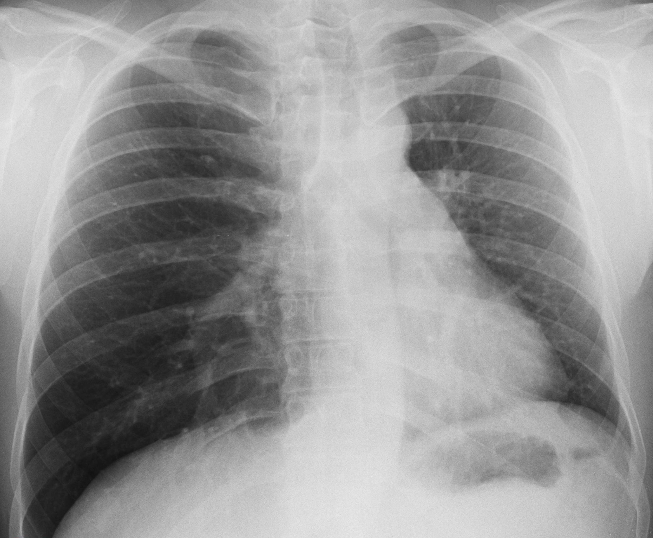
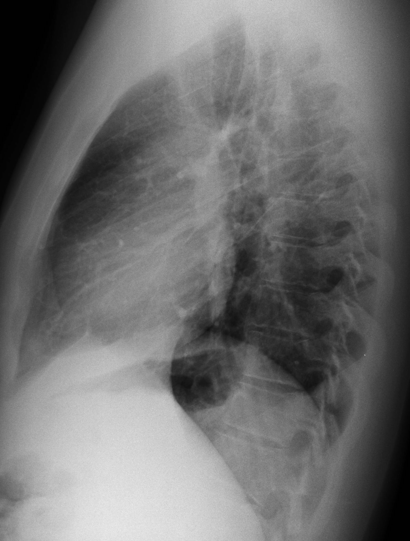
The main findings are seen in the PA radiograph, which shows displacement of the mediastinum towards the left. There is a discrete downward displacement of the left hilum (A, red arrow), as well as rotation of the cardiac silhouette causing loss of concavity of the left heart border (A, white arrow), the so-called flat waist sign, suggestive of LLL collapse. The findings are more evident when comparing with a previous film (B) taken two years earlier.

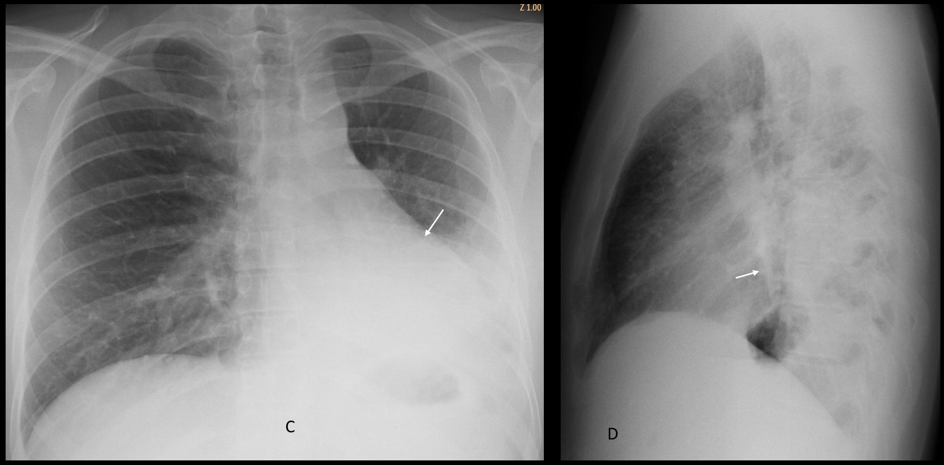
Findings were overlooked, and the study was read as normal. Five months later the patient returned with acute dyspnea. Chest radiographs show obvious signs of LLL collapse (C and D, arrows).
Unenhanced CT images show a rounded tumour in the left main bronchus (E-G, red arrows), confirmed to be a carcinoid tumour.
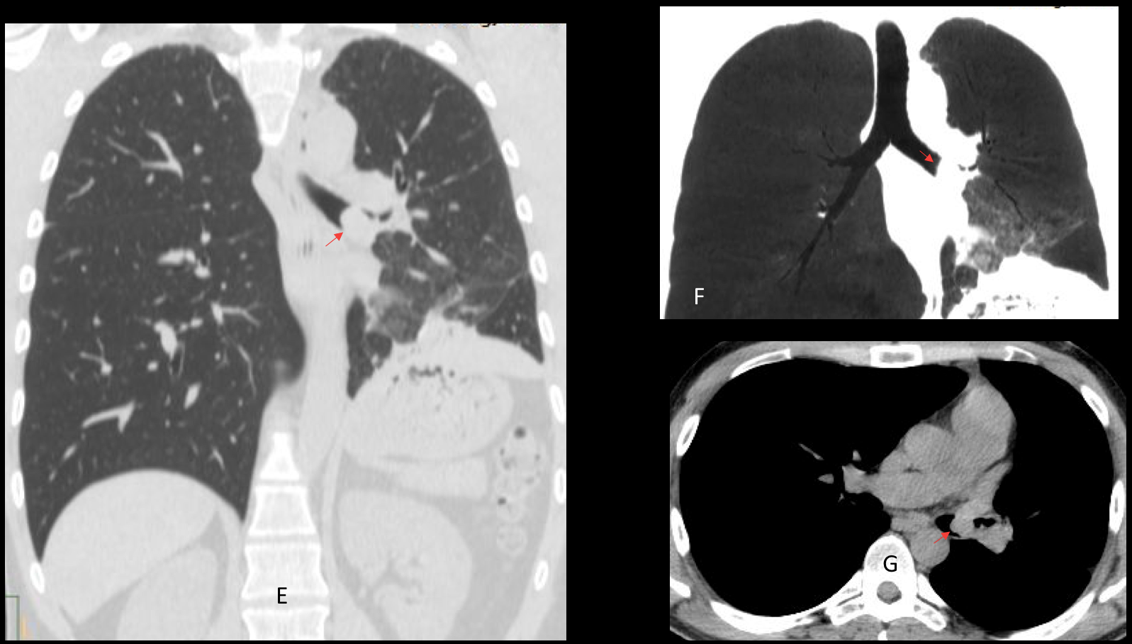
Final diagnosis: endobronchial carcinoid causing LLL collapse.
Congratulation to Beata who was the first to suggest the correct diagnosis.
Teaching point: remember to analyse the radiograph carefully in order to detect significant findings. In this case, several of you missed them and placed the abnormality on the right side, instead of on the left.


Left lower lobe collapse.
Greetings,
downward displacement of the right hemidiaphragm and mild shift of the mediastinum to the left; with hyperlucent right lung, all suggest right lung hyperinflation.
DDX: obstructive bronchial lesion (foreign body, tumor, external compression), bronchiolitis obliterance,… .
It is an interesting comment. Only problem is that you cannot be sure of your diagnosis unless an expiratory film is obtained 🙂
Scimitar sy
Remember that congenital pulmonary hypoplasia with scimitar vein from a practical point of view only occurs on the right side.
Uncross your fingers and try again 🙂
Hello. I think the right lung is abnormally hyperinflated and there is paucity of the vasculature. My first hypothesis would be Swyer-James.
See the answer to tr, above
Hello!!
The right hemithorax is overinflated and there is left mediastinal displacement towards the left side. A focal increased density next to the right hilum could be a mucus plug … so Congenital bronchial atresia will be a good option…??
No 🙂
left hilar enlargement,trombosis or oclusion of the right pulmonary artery
Gentilissimo Prof…..può essere il risultato di una malformazione adenomatoidecistica, la formazione in sede paracardiaca basale dx?Non ti dimentico….
Welcome, old friend! Your diagnosis is not correct. Sorry 🙂
The left hemithorax is smaller with ipsilateral mediastinal shift. Also there is a vertical tubular structure seen which most likely represents mucoid impacted bronchi. Could be due to bronchial carcinoid or some other intrabronchial etiology.
Good! You reconsidered your diagnosis and came back with the right diagnosis.
Congratulations!
Thank You for education!
Good! You reconsidered your diagnosis and came back with the right diagnosis.
Congratulations!