Dear friends,
Showing post-operative radiographs of a 34-year-old male. Muppet saw the films without clinical information and was completely lost.
Can you interpret the findings and shame Muppet into early retirement? Check out the images below, post your thoughts and possible diagnoses in the comments section, and come back next Tuesday for the answer!
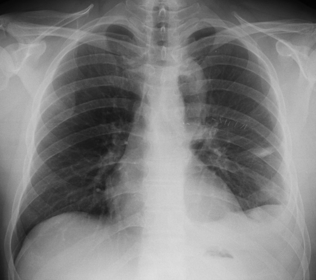
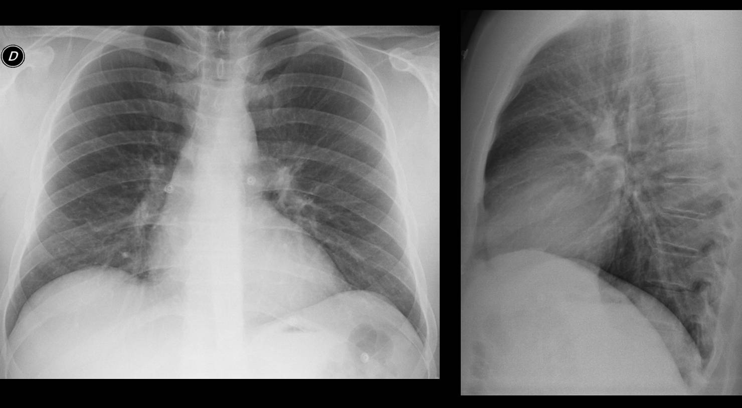
34-year-old male, PA chest
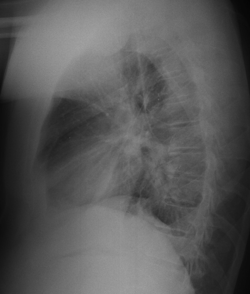
34-year-old male, lateral chest
Click here for the answer to case #72.
Findings: chest radiographs show post-operative changes in the left hemithorax, with surgical clips in the mid-field and residual pleural effusion. There is a para-mediastinal tubular structure, better seen in the immediate post-operative film (A, B, arrows). As the majority of you mentioned, a tubular structure in the lungs suggests a vascular lesion. Muppet investigated the clinical history and it corresponds to a graft between the subclavian artery and descending aorta in a patient with aortic coarctation. The surgeon found a hypertrophic Adamkiewicz artery with marked collateral circulation and was afraid to resect the coarctation and cause damage to the spinal cord (thus the graft).
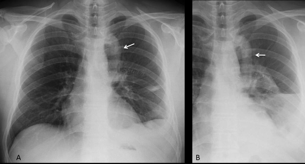
Fig. 1
Muppet checked the pre-op films (Fig. 2) and there were no imaging findings suggestive of coarctation: no rib notching, non-prominent ascending aorta and unremarkable aortic knob. This normal appearance may be related to collateral circulation going through Adamkiewicz artery.

Fig. 2
Final diagnosis: aortic coarctation corrected with bypass graft.
Teaching point: tubular images in the lung should suggest three possibilities: vascular, bronchial impaction and herniated bowel loops.


I think these x-rays images shows postoperative left sided clips,incapsulated left pleural effusions,catheter /drenage/,cardiomegaly,deformation of the left hilus,and tubular Ro positive shadow projecting in the left lung near left hilus.DD:1.Aerophagy 2.Surgery intervention because of vascular malformation.3.Mediastinal mass.4.Lobectomy 5.Superponated shadow from outside the lungs 5.Pneumomedistinum etc.
Left lobectomy, overexpanded LUL, downward displaced left hilum
There must be a case of semilobectomy with downwards shift of the left hilum and a bit of left semidiaphragm elevation.The tubular shadow near the upper mediastinum may be an abnormal vascular structure. The concave shadow is due to some kind of focal fluid collection.
The radiograph shows surgical clips (anterior minimal thoracotomy), effacement of the costophrenic angle (anterior pleural effusion), thickening cissural, left lung volume loss and left diaphragm elevation (segmentectomy vs atelectasis).
post operative lt subpulmonic effusion,surgical staples n a mostlikly drainage tube clearly seen in lat view for drainage of effuion?
I’m still trying to figure out the kind of procedure this patient had. The surgical clips are in a bizarre location (in a line on AP view and projecting on the vertebral column on lateral view). There are also round and tubular positive shadows near the downward shifted left hilum extending in the LLL, more easily seen on the lateral view (of vascular origin?). The weird tubular shadow near the mediastinum is starting at the level of the left clavicle and is extending over the left hilum with a hilum overlay sign up to the level of Th7 Th8. I am not sure I’m seeing it on the lateral view ( maybe projecting over the vertebral column and the middle mediastinum).
Hope I am not getting too close because I don’t want the Muppet to retire !!
I think I can see air filled tubular structures arising from the trachea at T3 level on either site, ? supernumeri bronchi and i am just wondering if the left sided tubular structure is just collapsed lung aerated by the extra bronchus on the left. crazy???
I think Muppet is here to stay and we don’t allow any thoughts for early retirement!!!!!!!
ps. Next Tuesday is way too far!!!!!
Sorry, Muppet is very strict about deadlines!
Muppet is human and is planning a two-month vacation after posting next case (and answer to this one, of course).
thoracotomy? left sided pleural effusion. Like post op? left paratracheal hollow shadow. Oesophagectomy and gastric pull through? but age is only 34… I wonder why gastric pullthrough in a 34 years old… Well! we will see… 🙂