
Dear Friends,
Muppet is in a good mood today (Miss Piggy may be responsible for that) and wants to present a ‘wysiwyg’ case (what you see is what you get).
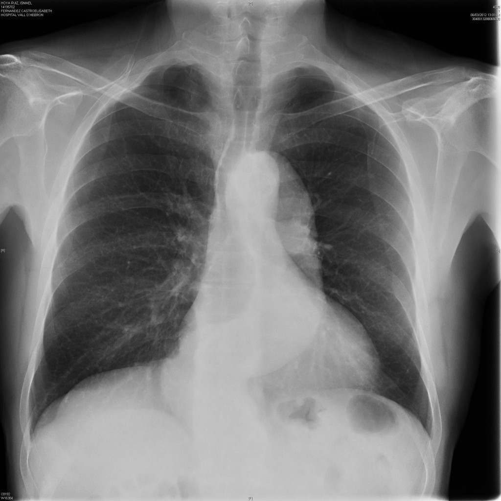
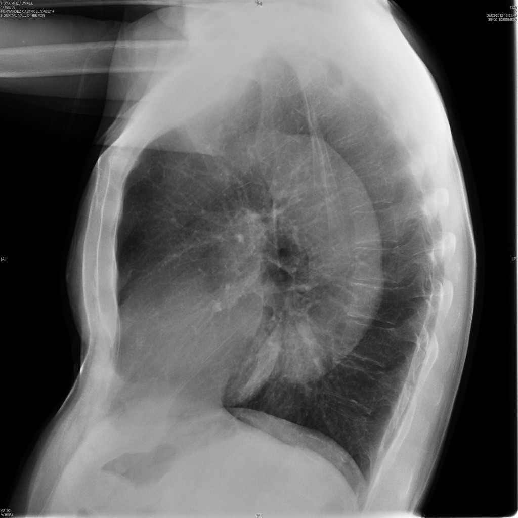
64-year-old, asymptomatic male. Chest images taken one month after an episode of myocardial infarction.
Diagnosis:
1. Aortic aneurysm
2. Aortic elongation
3. Type A aortic dissection
4. Type B aortic dissection
Clicke here for the answer to case #18
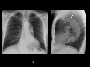
PA and lateral chest show an elongated descending aorta, while the ascending aorta is not prominent at all, as evidenced in both views.
A non-dilated ascending aorta goes against type A dissection and aortic elongation (whole aorta should be prominent). This leaves two possibilities: type B dissection and aneurysm of the descending aorta. In Muppet’s experience, the combination of a non-dilated ascending aorta and dilated descending aorta is highly suggestive of type B dissection. The transition from normal to dilated aorta can be seen in the lateral view (arrow).
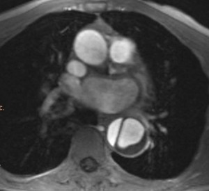
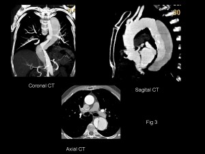
In this patient the dissection was discovered accidentally in a cardiac MRI performed to evaluate the heart after the myocardial infarction (fig 2). Enhanced CT confirms the diagnosis of type B dissection (fig 3).
Congratulations to all of you that made the correct diagnosis, except if you are supporters of Chelsea FC. Muppet is a Barcelona fan and hates losing!
Teaching point: enlarged descending aorta with normal-looking ascending aorta strongly suggests type B dissection, even though the patient is asymptomatic. Enhanced CT should be performed to investigate this possibility.
Also, never play a football match with a defence of three!


I would go for elongation and very likely aneurysm and do a ct. You can not rule out a dissection without contrast… ??
Can it be a marfan syndrome with aortic aneurysm of the descending aorta?
Marfan’s usually involves the ascending aorta
2. Evidently there is a aortic elongation.
The retrosternal space is clear, so, there is any ascending aortic aneurysm.
In the AP projection I am not sure to identify both margins of the descending aorta, so it make me difficult so say if there is an aneurysm. I need a CT
Performing an enhanced CT is the only way to have a definite diagnosis. But it’s more cool to suggest a possible diagnosis with the plain film.
l’aorta discendente è tutta dilatata proseguendo con un kinking nel tratto addominale : si veda la netta differenza di calbro in LL, tra,aorta ascendente e quella discendente.SI veda in ap un arco normale e la netta impronta-dislocazione della trachea. Non ci sono segni di scompenso del piccolo circ olo ed il paziente è aSINTOMATICO. lA DIAGNOSI PIù PROBABILE è DI ANEURISMA AORTCO-ADDOMINALE( TIPO c CLASS. safi).UNA ANGIO-TC CON RICOSTRUZIONE IN 3 D, DOVREBBE CONFERMARE LA DIAGNOSI.
The correct answer should be A. aortic aneurysm (the aorta is dilated as shown better in lateral view)
C + D should be excluded on clinical grounds (asymptomatic patient)
What if the dissection happened a few months ago?
A type A dissection would require some kind of therapeutic intervention which is not apparent here. So if that is case it should be a type B dissection.
Another remark is the presence of a moderate pectus excavatum (better appreciated in the lateral film) which can be related to Marfan syndrome and vascular compromise.
Looks like aortic dissection type B.
http://emedicine.medscape.com/article/416776-overview
Aortic elongation
Type B aortic dissection – maybe a PTA complication? He probably had one in myocardial infarction episode.
ANEURISMA AORTICO TORACO-ADDOMINALE: TIPO C SEC. SAFI. UNA ANGIO-TC, CON RICVOSTRUZIONE IN 3-D, CONFERMA LA DIAGNOSI.
i think it is aortic aneurysm, i cannot see the signs of aortic dissection by X-ray like calcium sign. widening of the mediastinum, pleural or pericardial effusion and i can determine the wall of the aorta well on contrary to the dissection.
NB are you sure that Muppet in good mood with this case.
Muppet is in excellent mood! (perhaps miss Piggy was a good influence). Happy to see you all sending answers.
He wishes to give all of you a diagnostic pearl.
Can someone please post the answer for this case?
Looks a little like an old aortic coarctation because of the step from arcus to descending aorta – and aortic aneurysm with or without dissection (only type B seems to be realistic, if even…)- here I need a second method, e.g. CTA or MRI. Dissektion as complication of PCI would be an type A most of all.
Ok back to the beginning: I guess there are signs for type B dissection – double aortic contour on level of aortic knob, and unusual placement of atherosclerotic plaques on distal descending aorta.
Type B aortic dissection
Dissection type A…Because we can clearly see the silluete of the aorta, and the silluete of the dissection maybe all over the thoracic aorta.
I think there can be two pathologies at a time a well, i.e aneurysm along with dissection but i ll go with aortic aneurysm since there has to be one choice.
It looks like type B aortic dissection,as here is a sharp widening and a thin line is present at the point of widening.But asymptomatic patient suggest more towards aneurysm.
For an aortic dissection I would like to see the displaced intimal calcification, but in this case i don’t see it.
The X ray, show an enlarged and elongated descending aorta
Aortic dissection Stanford type B. Intimal calcification flap is visible on profile at the superior limit of aorta at the site of the change in caliber. Sinuous descending aorta. Pectus excavatum.
Good. I was waiting for you. Better late than never!
I would go for Type B Aortic Dissection for all the reasons that have already been mentioned. Then I remembered a friend of yours with “abnormal” mediastinum on the plain film due to pectum excavatum ( ECR 2012 ). So I got confused!
Pectus will give an abnormal cardiac silhouette , but should not alter the aortic contour.
Type B dissection (the lateral view shows an indentation in the begining of the desending aorta), maybe an aneurysm, a contrast CT is needed.
check this link, birkin bag hermes price online JrxVxrgX http://www.hermesbirkin-price.org/
Pectus excavatum? Isn’t it pectus carinatum?