Dear Friends,
Muppet gratefully acknowledges the contribution of his good friend Dr. Manel Escobar who discovered and diagnosed the following case:
An 11-year-old girl with migrating bone pain for the last year, with normal radiographs, now presents with one month of low-degree fever. Chest and abdominal CT were unremarkable, except for the pelvic findings.
Diagnosis:
1. Fibrous dysplasia
2. Langerhans cell histiocytosis
3. Lymphoma
4. None of the above
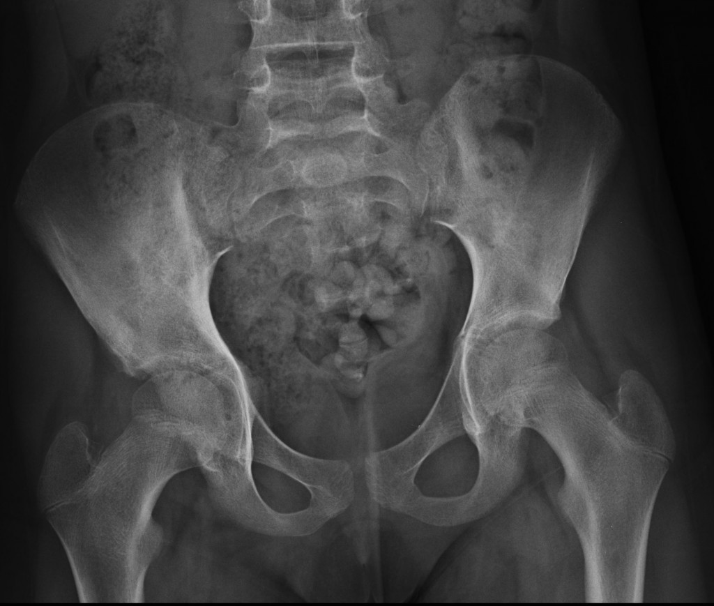
11-year-old girl, AP pelvis
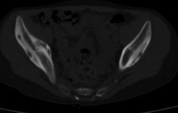
11-year-old girl, axial CT
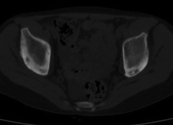
11-year-old girl, axial CT1
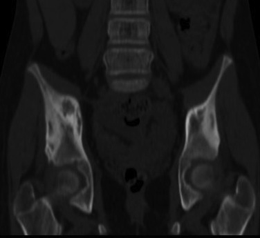
11-year-old girl, corinal CT
CT shows lytic lesions in both iliac bones with a thick periosteal reaction. A sequestrum is seen in the left side (arrow). The presence of a sequestrum and thick periosteal reaction suggests a chronic osteomyelitis. The clinical and imaging findings in this child fit an uncommon disease called chronic recurrent multifocal osteomyelitis (CRMO), which was the final diagnosis in this case.
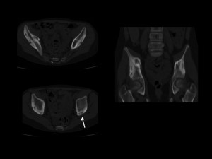
CRMO is an inflammatory process, which affects children. It is a nonbacterial osteomyelitis and typical imaging findings include lytic and sclerotic lesions of bones. Radiologists are usually important in determining the right diagnosis. In this particular case, findings were discovered by the radiologist and the correct diagnosis suggested.
Teaching point: Muppet is not an expert in bone imaging and does not dare to give advice. But he learned a lesson: from now on, chest imaging only!


Langerhans cell histiocytosis…I think…
Le lesioni sono addensanti: nel loro contesto ci sono aree litiche , ben definite e non ilfiltranti.La malattia è cronica, con febbre. Ecludere allora il linfoma.Escluderei la displasia fibrosa poliostotica.Rimarrebbe la istiocitosi a c. di Langerhans : ma sapendo le trappole del mio illustre collega, formulerei una diagnosi, inusuale per sede ed eetà: la M. di Erdheim-Chester
Muppet does not cheat (trappole). He just distorts reality…
No. 4 -none of the above; possibly CAFFEY – SYNDROME
It is not Caffey’s, but you gave an intriguing diagnosis. Muppet did not think of it.
No. 4. I would want to rule out Myelodysplastic Syndrome and/or Leukemia.
Multiple, radiolucent, cortically centered iliac lesions. They appear with reactive cortical thickening and sclerosis around them.
One of the lesions (left iliac in the 3rd image) seems to contain “bone inside bone”, it suggest a sequestrum. Only a few lesions can produce a sequestrum; classically, as far as I concern, basically 4: OSTEOMIELITIS, LYMPHOMA, EOSINOPHILIC GRANULOMA and some types of sarcoma (FIBROSARCOMA).
With this, the only one of these four differentials that could go with the case (even atipically) is MULTIPLE EOSINOPHILIC GRANULOMA (3.Langerhans Cell Histicytosis).
No more ideas by the moment…
Answer: 1
This is most likely polyostotic fibrous dysphasia associated with McCune Albright syndrome.
There are bilateral predominantly right pelvic Lucent lesions with associated sclerosis and bony expansion noted.
Coxa vara.
Accelerated bony maturity.
This is associated with precocious puberty and bone pain may be due repeated fractures
This is more common in young girls.
I think that it is fibrous dysplasia as a part of MCcune Albright syndrome
you will like cheap burberry outlet , just clicks away
for coach wholesale at my estore RGMoNmcf http://www.wholesalecoachhandbags.us/