Muppet has chosen to show a case from the Iron Islands. Winter is coming …
An 83 year-old lady with cough and moderate dyspnea.
1. Normal for age
2. One process
3. Two processes
4. Three processes
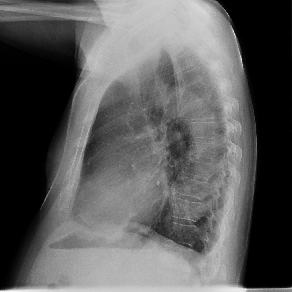
In the PA view there is an intrapulmonary nodule in the right costophrenic angle. It is intrapulmonary because the lower border is surrounded by air (arrow). In addition, the descending aorta is prominent, whereas the ascending aorta is not. This is a sign which suggests type B aortic dissection in the plain film, which should be confirmed or discarded by CT.
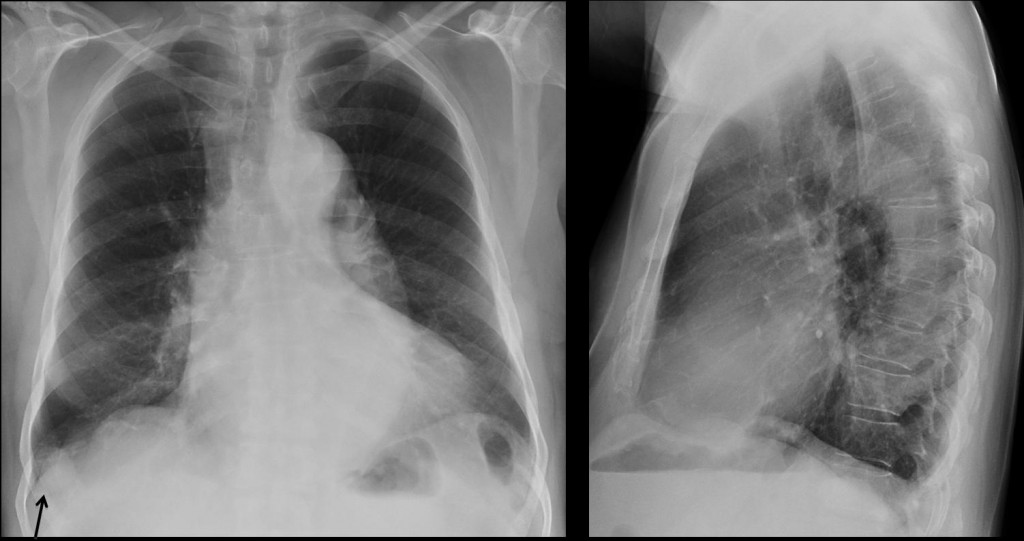
Fig. 1
CT confirms the dissection (A, B arrows), as well as a low-density nodule (15 H.U.) at the right base (A, C arrow). Nodule had not changed at one-year follow-up. Final diagnosis: two processes, benign lung nodule and type B aortic dissection.
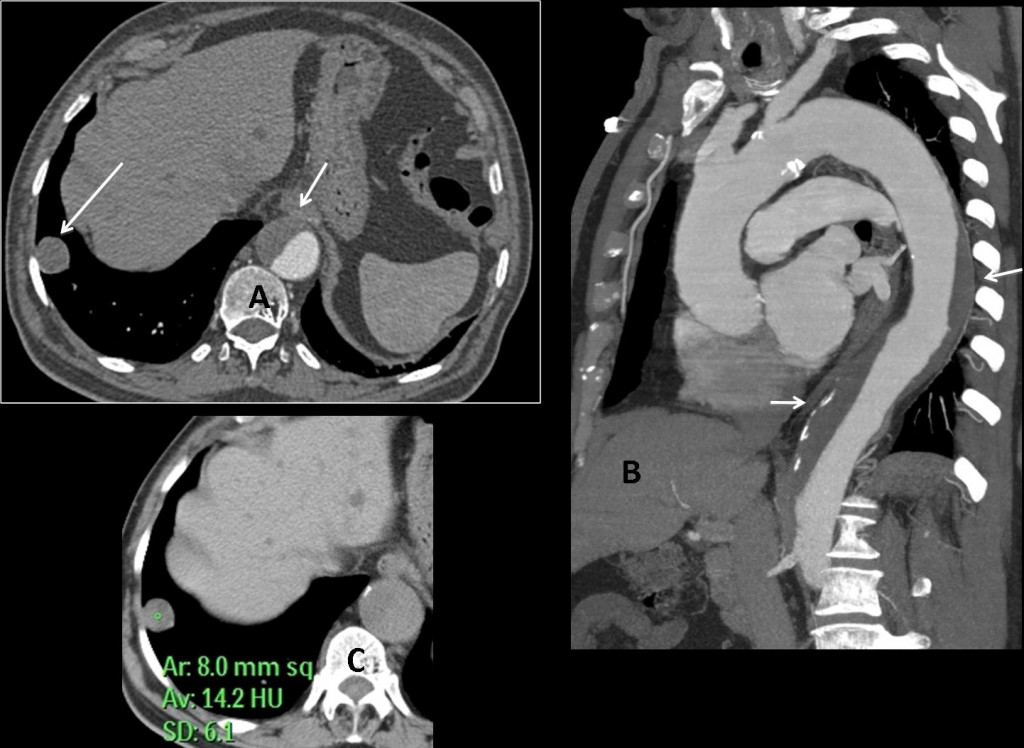
Fig. 2
Teaching point: always look at the costophrenic angles in the PA chest. You may find a lesion occasionally (as in this case). Remember that when, in old age, aorta elongates, there is prominence of both the ascending and descending aorta. When only the descending is prominent, discard type B dissection (see case 18).
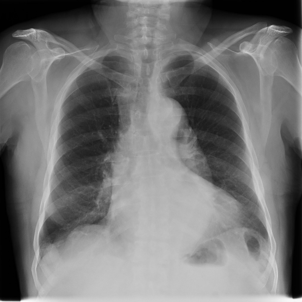


What happened to Muppet fans? Have they deserted? Is Dr. Pepe more atractive? Or you find the case too difficult?
Since I cannot detect any other abnormality, I will just comment on the heart size and aortic unfolding, which are not surprising at this age. But I suspect that is not why the case is up.
So, you choose number 1 ( normal for age). Let me warn you that
in my dilated life I have never seen Muppet showing a normal case
Si può cambiare moglie, si può cambiare religione ,ma non si può cambiare il tifo per la squadra del cuore: rimango fan” dell’illustre collega! Il caso presentato è insidioso:quello che mi sembra vedere è in LL ove l’immagine cardiaca sembra mostrare una tenue formazione rx-opaca sul profilo cardiaco inferiore, che sembra improntare la bolla gastrica.Potrebbe trattarsi di unao pseudo-aneurisma cardiaco, esito infartuale.
la signora è stata mastectomizzata a sx.
Sorry, my mistake. Is a 78 y.o. male. And the case is certainly “insidioso”. Keep looking
There is a mass-like lesion in the Region of the right lateral pleural sinus (I am Not sure where it is on the lateral View – i Hope it is not the nipple again?!
The Left lung looks slightly more transparent than the right one.
Other than that i have no clue. Very difficult case – muppet have mercy 😉
En la proyeccion frontal se identifica una opacidad de contoneos definidos localizada en el contorno del cardiomediastino izquierdo compatible con masa, en la proyeccion lateral no se define muy bien. Y también creo que puede ser un pseudo aneurisma cardiaco.
Right-sided diaphragmatic eventration – partial?
Contours of the right hemidiaphragm is double wavy.
or is it a homogeneous round mass above the right diaphragm, two, one larger and one smaller laterally. It is located on the lateral projection of the summation of the heart shadow……Hydatid Cyst Of The Diaphragm? 🙂
Close enough. Anything else? Or you choose answer 2?
Coud we have a beat big images?
The sight of some ancient residents as Lola is becoming less and less eaple-eyed if sometimes was it.
Please, don´t make to much jokes about the glasses or the age.
What you have to see is big enough. Muppet have a 27″ Mac
and sees it very clearly.
Hmm just a wild guess:
There seems to be a double contour of the aortic arch and of the descending aorta.
Is this real?
Go on.
It is obvious that there are two lesions, perhaps mother and daughter cysts, perhaps spread through the diaphragm from the liver to the lungs. Larger cysts have thicker walls and smaller mimics soft tissue mass ….. different stages of hydatid cysts?
hm … diff.dg. but less likely pulmonary sequestration.
If the patient has had previous trauma, then it could be a partial rupture of the diaphragm with herniation …..
3. Two processes and …definitely a CT scan …
CT coming shortly
per il doppio contorno dell’arco aortico ed aorta discenente si può pensare ad una dissezione aortica tipo B.
Excellent!
I see two homogenous round shadows against right diaphragm-smaller one laterally and bigger one(its cranial outlines) medially.In the lateral view they are digned agains the heart shadow – lesser seen better.
I am not so sure but in the lateral view against Th12 I see another one shadow??Is this the contour of left diaphragm – I am not sure?
and if you count last… my answer is 4(how brave:) )
Perhaps too brave…
I know,so if I have to be honest I am sure for two processes- one rounded shadow against right diaphragm(laterally) and dilated outlines of the descendent aorta.
FOR ME ITS 2
Old patient plus enlarged descending aorta, I think the first option could be an aortic dissection type B.
Furthermore ,I wonder about the acygos vein in the frontal view…could be a little enlarged??
Perhaps azygos is prominent, but has no significance in this case. Good diagnosis
1-COPD
2-cardiomegaly
3-right space occupying mass in segment 10.
please don’t keep me waiting for the answer or I will end with 35 processes
Sorry,you will have to wait a little longer. Combine your answer with Marcy above
I think the old lady’s x-ray shows two processes:1.Interpositio colonis. 2.Pectus excavatum
To prove Ao dissection- CT with contrast.
Great discussion! I guess i’ll go for three processes:
1- Cardiomegaly;
2- aortic dissection type B;
3- right space occupying mass(es) in the middle lobe (with partial lobe atelectasis maybe?)
i think it is a pathalogy in retrocardiac space
– may be in the descending thoracic aorta, aneurysm or disection with some atherosclerotic changes.
-we have also to exclude esofageal pathology , like achlasie or neoplasm.
-pulmonary lesion must be excluded like a colaps which asociate elevation of the left hemidiafragm and traction of the left main bronchus.
The right Hemidiafragm have a boselated contur but still there are 2 or three round lesions under the right diafragm, 2 anterior and one posterior which may be hydatid cysts in the liver .
one process
one process unfolded aorat
3 processes. The unfolded aorta with a double shadow, maybe an aneurysm, the right diaphragmatic eventration and the prominence of the central pulmonary vessesl with early pruning.
Can we have larger images?
Thanks for the cases – very useful.
If you double-click on them, they will get larger. Thanks for your opinion
BTW – I think the most important findings are the one mentioned by the guy from Bari, together with loss of the aorto-pulmonary window.
Siamo ai tempi supplementari e al “golden -Share).1= DISSEZIONE AORTICA TIPO b. 2=ESITO INFARTUALE CARDIACO, CON PSEUDO-ANEURISMA.3 cISTI CELOMATICHE DEL PERICARDIO( A DX).4= ADIPOSO-MASTIA DEL SENO DX.
I agree with the hypothesis of dissection of descending aorta .Should I add to this, the presence of dilated LT subclavian artery and pulmonary veins.I also vote for 1 liver lesion.The outer lesion in the ap xray has a radiopaque periphery.(maybe a skin lesion?).I also agree with the presence of adipose tissue in the RT breast.
the solution is by dilatacion aneursimatic type B AND BENIGN NODULE
you must read dallas cowboys jerseys for less
It looks mural thrombus rather than disection on CT. The filling defect is seen internal to the intimal calcification.
Can you please opine on this ?