
Dear friends,
Welcome back to Caceres’ corner.
I’m already keen on your suggestions on Case #4.
Clinical history: Pre-operative chest radiograph for ophthalmologic surgery in a 57 y. o. male
Most likely diagnosis:
1. Carcinoma of the lung
2. Unilateral hyperlucent lung
3. Pulmonary embolism
4. Giant bulla

Pre-operative chest radiograph for ophthalmologic surgery in a 57 y. o. male (PA chest)
Click here for the answer to case #4
My initial impression of the PA chest is a left hyperlucent lung with diminished vascularity. Muppet is smarter and always looks for other findings. He sees a very low left hilum (red arrow), a paraspinal oblique line (blue arrows) and blurring of the left hemidiaphragm on the lateral view (white arrows). These findings are very suggestive of left lower lobe collapse. The most common causes of collapse of the lower lobes are bronchiectasis and carcinoma. Considering that the former was not included in the options, the most likely diagnosis is carcinoma of the lung.

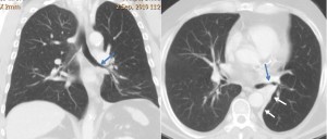
Diagnosis is confirmed with CT, which shows irregular narrowing of the bronchus (blue arrows) and marked collapse of LLL (white arrows). Biopsy: epidermoid carcinoma.
Teaching point: remember case #1. Always look at the hila. They are very useful markers in lobar collapse. Muppet will be very disappointed if you do not remember this!


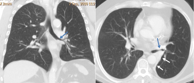


Left loewr lobe collapse most probably due to bronchogenic carcinoma
– hyperlucent left lung with soft tissue density at the posterior costophrenic recesss may represent giant congenital upper lobe bulla causing compressive atelectasis of lower lobe.
– hilum appears little prominent on lateral view. although unlikely, hilar lesion causing ball valve obstruction of upper lobe bronchus with obstructive emphysema needs consideration.
needs CT for furthur evaluation.
Yes, but what is your preferred diagnosis?
for the benefit of patient, i would like to rule out hilar lesion and recommend CT.
Personal preference would be giant bulla
not sure to include Mcleod’s in the differential.
looks like a giant bulla. Wondering about the hilar prominence though!
Goitre
Carcinoma of the lung.
Lower left lobe atelectasia due to hiliar located lung cancer
There is lower displacement of left hilum, as well as “sail sign”, all suggestive of LLL atelectasis, and fro given possible DDx, I would choose carcinoma of the lung
Giant bulla!
1. Carcinoma of the lung
Nice case,very interesting…facts:
Atelectasis cousing elevation of the left diaphragm and dislocation of the mediastinal structures ipsylateraly-there is no signs of any of it.
Left hemihorax is evidently hiperlucent.
No signs of any compresion of the vascular structures causing by bulla.
Left hila is in litlle beat cranial position,and left mean bronchus is more verticaly oriented.
Right hila is more vascular then left.
Paravertebral shadowing on the left-meaning pathology in azygooesophageal recess which is widened?
Verry nice,enough facts for MSCT 🙂
left hila is a in a litlle beat CAUDAL position…sorry,my mistake
Nice description, but I still need a diagnosis before the CT!
Well,Mr.Prof. my final decision is hiperlucent left lung,but there is some signs of three others DDx. in it…
LLL atelectasis
Most probable diagnosis : carcinoma of the lung
Well,Mr.Prof. my final decision is hiperlucent left lung,but there is some signs of three others DDx. in it
1. Carcinoma of the lung.
Could be to an endobronchial foreing body.
LLL colapse signs lead me to choose carcinoma of the lung as the most suitable diagnosis.
Giant bulla!
pulmonary embolism