Muppet is so happy with your performance that he has chosen a challenging case. The following radiographs belong to a 52-year-old woman with two episodes of chest pain in six weeks. The initial radiograph is shown, as well as a radiograph taken during the second episode. At that time, a CT was done.
1. Pulmonary infarct
2. Lymphadenopathy
3. Loculated pericardial fluid
4. None of the above
Answer to case 40
Initial PA chest shows a paracardiac mass (Fig 1A, arrow). In CT the mass is of fatty density, with a capsule that enhances after injection of IV contrast (B,C yellow arrows). There is mild pericardial thickening (C, red arrow).
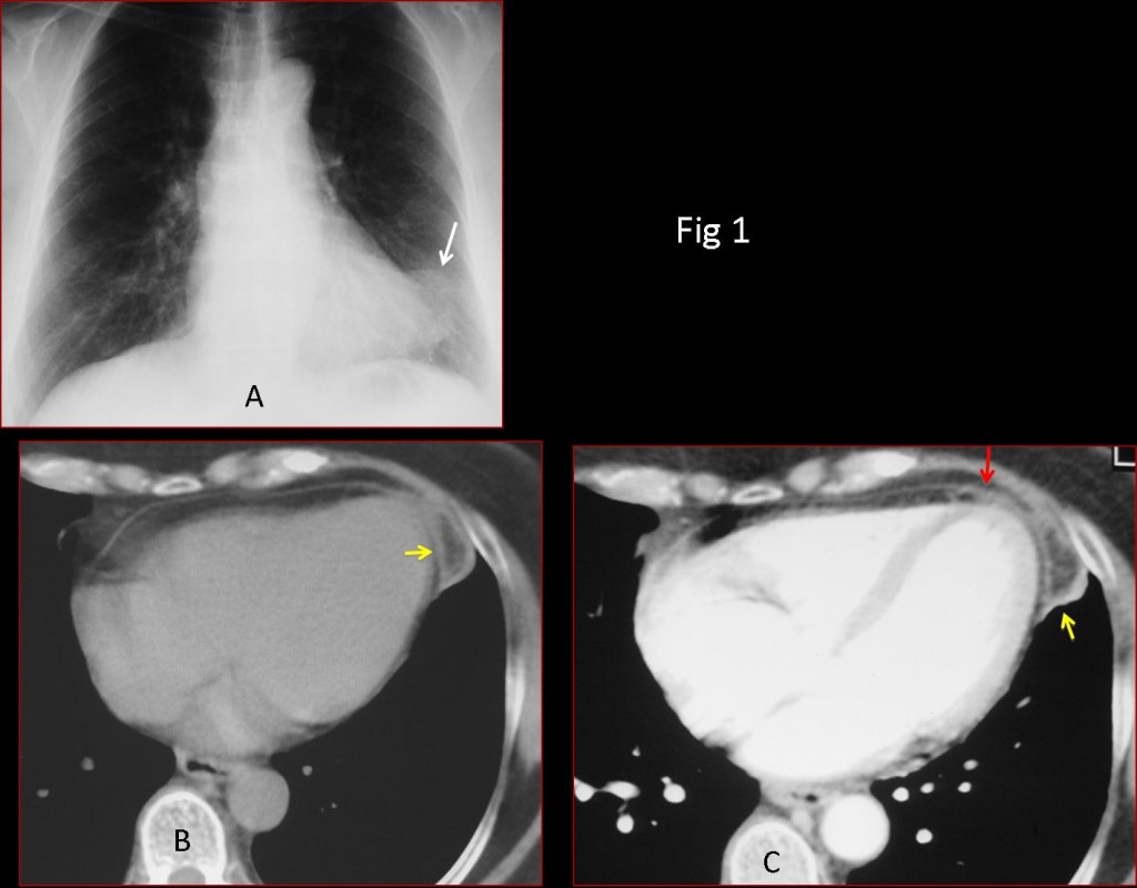
Fig. 1
The appearance is pathognomonic of epipericardial fat necrosis. Diagnosis is confirmed with follow-up imaging. The mass has shrunken markedly on CT (Fig 2B, arrow) and is no longer visible in the chest radiograph.
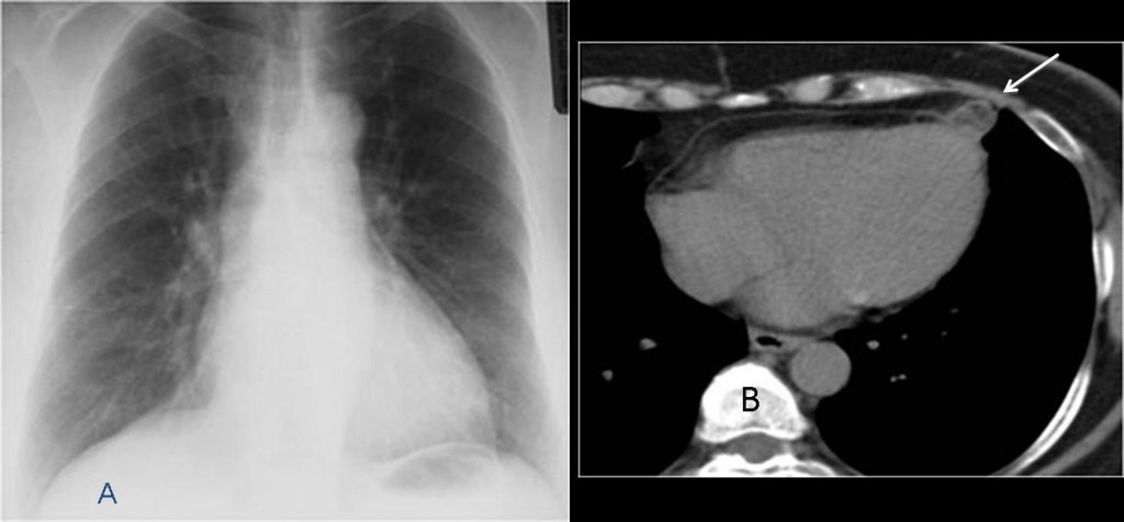
Fig. 2: Two months later
Final diagnosis: epipericardial fat necrosis.
Congratulations to Muppet’s faithful followers, who attained 100% correct answers (didn’t expect any less of you!). They may not know that this entity was described in the radiological literature in 2005. The initial description was made in 1957 and only 20 cases had been reported since; all of them operated on. Since 2005, this entity is being described with increased frequency (aside from isolated case reports, two educational exhibits at the ARRS meetings in 2011 and 2012 showed 10 and 14 cases respectively).
Teaching point: EFN is an easily recognisable entity, more common that believed and should be considered in the differential diagnosis of acute chest pain.
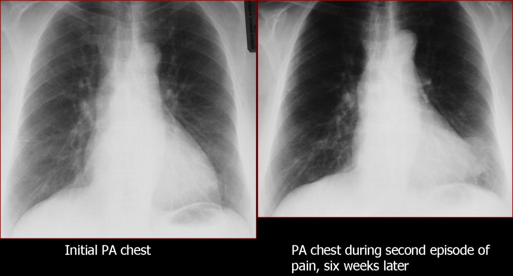
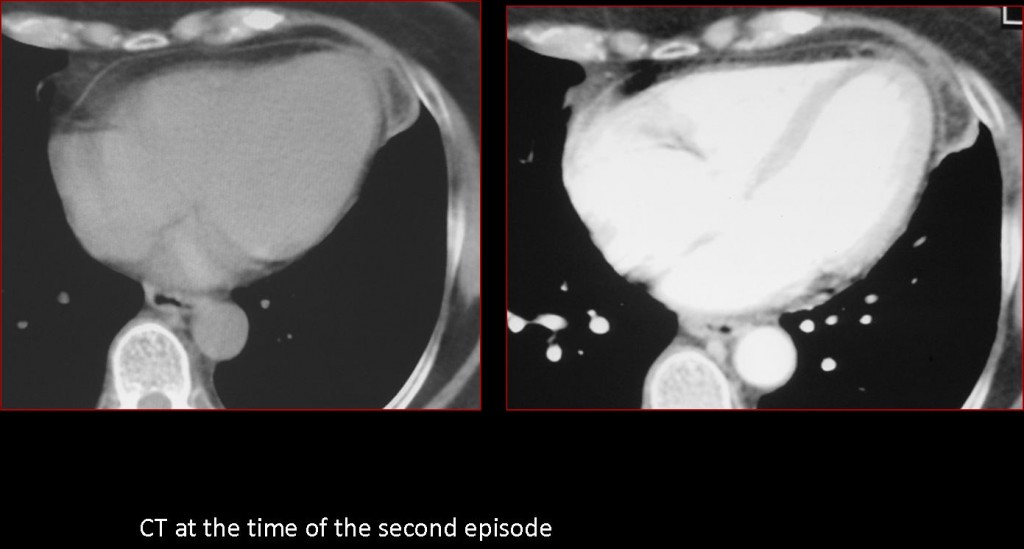


In the second Rx there is an abnormal convexity of the apical heart shadow on the left. On the CT there is a pseudonodular incresed attenuation of the epicardial fat next to the heart apex.
I would suggest an epicardial fat infarct (somebody call it epicardial appendagitis but I think it is equivocal).
I agree with albert. Also known as epicardial fat necrosis.
Pericardial fat necrosis
The chest film identifies an area of increased opacity in the region paracardiac left. In axial CT identifies injury margins encapsulated fat crisp uniform.
In this context clinical findings are compatible with pericardial fat necrosis.
You are too fast guys 🙂
Agree
Too fast indeed! Pericardial fat necrosis! 🙂
PS – Dr Caceres it was great to hear your lectures one week ago in Barcelona!
Good! Glad you remember. At least one person was not sleeping
Pericardial fat pad necrosis
Forse l’illustre collega è troppo buono ed euforico per il 5 a 0 del Real al Maiorca, per cui ha proposto un caso x far fare bella figura ai concorrenti: necrosi appendice pericardio.
there is a lesion of fatty density that abuts the pericardium of cardiac apex. After the iv contrast administration there is only minimal periferal enhancement. If we combine these findings with the clinical history of pain , it could be a rare focal epicardium fat necrosis, although we can not see the fat <> at the first radiograph.
We have to consider other possible diagnoses too. For example an epicardiac cyst , with transient exudative inflammation , explains the pain ,the nearly fatty and the flecs of soft tissue density, why we can not see it at the first exam, and the contrast patern.
As regards the loculated pericardial fluid , it could be such a case , but we would have expect to see clues of chrocic pericarditis, which we don’t.
This is not a common site for lymphnodes. The rare epiphrenic lymphnodes , usually happen at lower positions.
And of course this is not the classic appearance of a pneumonic infact.
Since 2005 I have seen seven cases of pericardial necrosis in Barcelona. I believe this entity is more common than believed.
And some cases present with a normal chest radiograph, They are discovered with CT.
Congratulations for an excellent discussion
Thanks a lot for your beatiful and educacional cases, Dr. Caceres!
Thank you. That was a nice thing to say.
For the diploma examination ,written part, Which boks should we use?
Good question. Will ask the persons in charge to find out if they have a recommended list.