Spotlight on radiology in Uganda

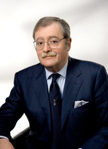
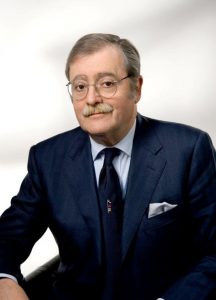
Michael Grace Kawooya is a Professor of Radiology at the Ernest Cook Ultrasound Research and Education Institute and Professor Emeritus at the Makerere University College of Health Sciences in Kampala, Uganda. He has done much for the development of radiology in his country and the rest of Africa, but says efforts must continue to increase the number of radiologists and range of equipment, and to raise awareness of radiation safety. Kawooya believes Africa can learn a lot from European advances. His contributions to improving bilateral cooperation will be rewarded today as he receives ESR Honorary Membership.
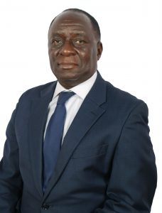
Professor Michael G. Kawooya’s contributions to improving bilateral cooperation between Africa and Europe will be rewarded today as he receives ESR Honorary Membership.

ECR Today: How much has radiology advanced in Uganda?

Michael Grace Kawooya: Back in the late 1980s, radiology was very new to medical practice in Uganda, and its contribution to healthcare was not well understood. It was not given priority and was underfunded. There were only two radiologists in the country when I finished my residency and we were overwhelmed with work. Doctors were not willing to undertake radiology residency, fearing that radiologists didn’t earn much. Equipment was scanty and often malfunctioning. Many of these challenges still exist today, but to a lesser extent. Radiology is better understood and its role is now evident. The number or radiologists has increased to almost 70. This year alone, 20 doctors took up radiology residency. The number and range of equipment has also increased.

ECRT: Are there any regional trends in radiology in Africa?

MGK: The same challenges facing radiology in Uganda bedevil most of Africa, but North Africa, which is largely Arabic, and South Africa, which is wealthier, face fewer challenges. In these parts of Africa, radiology has flourished more compared to Central, East, and West Africa. The radiologist-to-population ratio is approximately 1:67,000 in Egypt, 1:1,600,000 in Uganda and 1: 8,000,000 in Malawi. The more affluent regions have higher numbers and range as well as sophistication of imaging equipment. They have more radiology training institutions and undertake more research. Read more…

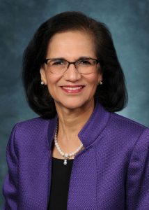
Ex-RSNA chief calls for more international cooperation

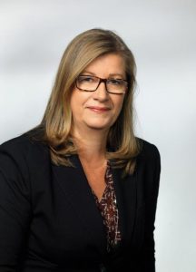
Vijay M. Rao, MD, FACR is the David C. Levin Professor and Chair of the department of radiology at Thomas Jefferson University. She is also senior vice-president of Enterprise Radiology, Jefferson Health, and the immediate past president of the Radiological Society of North America. As she receives ESR Honorary Membership today, she looks back on 30 years of work in a field she describes as globally united.

Radiology has come a long way from the days of plain film hypocycloidal tomography in head and neck imaging, a subspecialty to which Prof. Rao has dedicated most of her career. Technological innovations such as ultrasound, CT, MRI and PET have revolutionised the field, allowing for earlier diagnosis and greater insights into the pathophysiology of a host of diseases and conditions.

Professor Vijay M. Rao from Philadelphia, immediate past president of the RSNA, will receive ESR Honorary Membership today during the Grand Opening.

Radiology continues to evolve from anatomic to more physiologic and functional assessment of disease processes, with more precise quantification and minimally invasive therapeutic options.

Rapid technological advances have led to tremendous growth in radiology along with associated costs. But the digital age has introduced a number of previously non-existent opportunities to transform radiology practice, using tools powered by informatics and machine learning (ML). “Radiologists should explore ways to utilise these technological advances to add value and reduce waste in healthcare,” she said.

Artificial intelligence (AI) and ML applications are valuable tools that make radiologists more effective and increase their contributions to personalised and precision medicine. “AI can assist radiologists at a time when we are challenged to provide imaging services that are faster, safer and affordable, as well as information that is quantitative and precise. These new technologies will improve workflows in our daily practice, freeing up time for us to better position ourselves as integral members of the patient’s healthcare team,” she said.

The move toward value-based imaging continues to be the biggest trend in the US, where emphasis is placed on volume when structuring radiology practice. “Policymakers lack understanding of imaging’s contribution to patient care and the extent of services that radiologists provide. That is why we have begun the necessary transition to a value-based model of care, where radiologists are actively consulting with patients and their referring physicians as part of the healthcare team,” she explained. Read more…

Welcome to ECR 2019!

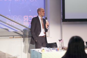
Welcome to the 25th ECR in Vienna!

Thanks for coming to be a part of ‘the bigger picture’!


By Lorenzo E. Derchi, ESR President

ECR 2019 marks an important anniversary. This will be the 25th ECR congress held in Vienna since 1991: a great achievement! Those who have been coming here since the beginning will find a meeting that has become bigger over the years, now including not only the Austria Center, but also many of the buildings surrounding it.

It is a true ‘congress city’, which has grown to accommodate the increasing numbers of delegates from all over the world. Those who are here for the first time will discover a congress with a wide range of opportunities to learn about the different aspects of our specialty, with sessions suited to all, from beginners to advanced professionals.

ESR President Prof. Lorenzo E. Derchi is Head of the Department of Radiology at the University of Genoa, Italy.

Over the years, the ECR has provided radiologists with a European platform for the presentation of research and debate and has been the key to the creation of a community of people working together and advancing in science and education together. This has been the result of the efforts of 24 Congress Presidents and Programme Planning Committees who, with the help of the well-structured and dedicated efforts of the ESR Scientific Programme Department, have worked as in a relay team, passing the baton from one to the next and ensuring continuity of aims and good results over the years.

The ECR has always been a creative meeting. It started with a ‘classical’ structure, made up of refresher courses, scientific presentations and posters, and, over the years, has incorporated various new ways for contributors to present scientific work. The first big innovation was in 2003, with the introduction of the electronic posters of EPOS. Then, in 2016, came the new session format ‘the Voice of Epos’ in which the scientific message of selected posters could be explained in person by their authors. Last year, the ‘MyT3’ presentations, the lounge meetings of ‘Coffee & Talk’ and the hands-on approach to interventional radiology of ‘the Cube’ were introduced. All these session formats have proven successful and will be continued, and even expanded, this year. Furthermore, an additional new session type will be launched, not only for ECR delegates, but also open to anyone who is interested in joining in. The ‘Women in Focus’ initiative will explore the challenges encountered by women in our field and in other medical and medical-related professions. It will open a debate over gender parity in our profession and will try to present role models to whom the ever-increasing number of women entering into radiology can relate. Read more…

Keep ultrasound close and generate added value, Lubinus advises

Professor Federico G. Lubinus is full professor of radiology and director of the radiology residency programme at the autonomous university of Bucaramanga (UNAB) in Colombia. He is one of the leaders in South American radiology and he believes that bringing back ultrasound to the heart of radiological practice is key to the future growth of the specialty. His efforts will be acknowledged today as he receives ESR Honorary Membership.

When it comes to radiology, South America has very similar challenges to the rest of the world and a number of regional trends, Lubinus explained. “Big workloads, turf battles with other medical specialties, and challenges imposed by teleradiology and artificial intelligence are some of the many difficulties we have to deal with, not only as radiological societies but also as radiologists wondering about the role of the specialty in the near future,” he said.

Professor Federico G. Lubinus, who will receive ESR Honorary Membership at today’s Grand Opening, is one of the leaders in South American radiology.

But radiology practice in the continent is uneven and a major difference concerns radiology training, according to Lubinus, who currently heads the Latin American Educational Forum, a group that brings together radiological societies of South America to develop and advance educational issues in the region. “Training is dependent on the regulations of each country. There is great diversity both in the number of years of study required to be a specialist and in the minimum requirements demanded in each country, which makes it difficult to standardise programmes and perform appropriate evaluation of knowledge,” said Lubinus.

Lubinus is also director of the scientific committee of the Asociación Colombiana de Radiología (ACR), a society he served twice as president. Under his aegis, the ACR worked to have a standardised training programme, a tool that is now recognised by the Colombian ministry of education as the basis for evaluation and certification of radiology graduates, and radiologists who have received their certification abroad.

For the past four years, the Latin American Education Forum has also engaged in developing a standardised academic residency programme as a reference instrument for the validation of qualifications of Latin American radiologists, to improve the much needed certification and recertification in radiology.

Read more…

Russia’s preeminent radiologist continues to reach for the top, after two decades with the ESR

Professor Valentin Sinitsyn is chair of radiology and head of the radiology department at the medical faculty of Moscow Lomonosov State University, Moscow. A pioneer in cardiac imaging in Russia, he has helped advance the field in his country, but believes there is still much to be done to increase MR and CT use. After 20 years of working to advance the ECR and European radiology, he will receive the ESR Gold Medal today.

Sinitsyn has witnessed the introduction of spiral and multidetector CT, high-field MRI and hybrid imaging, and the development of evidence-based radiology and medicine.

Professor Valentin Sinitsyn from Moscow

Professor Valentin Sinitsyn from Moscow, a pioneer in cardiac imaging in Russia, will receive the ESR Gold Medal today.

As he graduated from Sechenov Medical University in Moscow in 1984, cardiac CT and MRI did not yet exist. “With the help of these new modalities, radiology penetrated fields where it had never been used before, and the best example is cardiac imaging,” he said.

Sinitsyn started implementing cardiac imaging back in the 1980s and has very fond memories from that time. Although these were also harsh days for the field, he said.

“Most cardiologists and radiologists believed that cardiac MRI and CT were toys that were good for nothing in real clinical practice. I still remember my first case of apical hypertrophic cardiomyopathy diagnosed with cardiac MRI in 1987, when nobody could make a correct diagnosis,” he said.

Today he admits to being a little disappointed with the slow penetration of cardiac MRI and CT in Russian clinical practice. The benefits of modern cardiac imaging are well known, but the percentage of cardiac CT and MR examinations in cardiac imaging is just around 1%. “Low reimbursement for such examinations and insufficient knowledge and motivation from both radiologists and cardiologists are mostly to blame for this situation,” he said.

Read more…

Incoming ESR President Lorenzo Derchi looks forward to celebrating an ECR milestone in 2019


By Julia Patuzzi

As has been our tradition for a few years now on the final day of our congress, we already look ahead to next year’s ECR. We therefore spoke with Prof. Lorenzo E. Derchi from Genoa, Italy, who, as incoming ESR President, is in charge of ECR 2019. He told us about his plans and ideas for the next European Congress of Radiology.

ECR Today: Professor Derchi, next year’s European Congress of Radiology will be the 25th to be held in Vienna. Are there any specific celebrations planned that you can already share with us?

Prof. Lorenzo E. Derchi

As incoming ESR president, Prof. Lorenzo E. Derchi, Head of the Department of Radiology at the University of Genoa, will preside over ECR 2019.

Lorenzo E. Derchi: We are still thinking about them. An anniversary like this is something more than an opportunity for a big celebration. It is the right moment to re-think our history and what we have reached over the years. To always have the congress in the same city since 1991, at first every other year and then, after 1999, annually, has been a radical change. The new formula proved successful and ECR has become the European meeting to come to in order to learn the latest clinical and technical advances in our discipline as well as to present the results of the previous year’s research.

In my experience, however, it has become much more. From 1991 up until today all congress chairpersons have been part of the same team in a relay race, each handing off the baton to the next one. This continuity can be felt in the special atmosphere at each meeting, and has allowed the congress to become the annual appointment to meet old friends and make new ones; to discuss any radiological topic and then to start working together on it; and finally a place to meet periodically to share problems, advancements and results of long-term projects.

ECRT: Were you here for the first ECR in Vienna in 1991? If not, when did you attend your first ECR? What do you recall from that visit?

Read more…

Trojanowska to deliver honorary lecture on neglected form of cancer

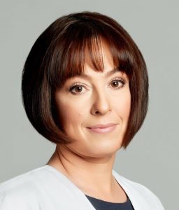
Polish radiologist Dr. Agnieszka Trojanowska will shed light on human papilloma virus (HPV)-induced squamous cell cancer of the head and neck, a common yet long neglected type of cancer. In an interview with ECR Today, she also explained the origins of her passion for head and neck imaging, and how radiologists will increasingly benefit from computer science.

Dr. Agnieszka Trojanowska, assistant professor in the department of radiology and nuclear medicine at Lublin University Hospital, is an internationally recognised specialist in head and neck imaging. Her special interest for oncology and her natural curiosity led her to agree to deliver the Josef Lissner Honorary Lecture on HPV-induced squamous cell cancer of the head and neck (HNSCC) at ECR 2018.

Agnieszka Trojanowska from Lublin, Poland, will speak on HPV-induced squamous cell cancer of the head and neck in her Honorary Lecture today.

“HNSCC behaves differently than more well-known forms of SCC, and it is becoming more and more prevalent. It affects young, well-educated people with high socioeconomic status, and is a quickly rising sexually transmitted entity with peculiar clinical and molecular characteristics. In particular the rise of HPV-induced cancer has been observed in the United States since the beginning of 21st century,” she said.

It was important for her to talk about this long overlooked topic, for which much remains to be done. “It has been under-estimated for many years in many countries. Nowadays, we can speak about an HPV epidemic, leading to a significant rise of oropharyngeal cancer incidences worldwide. These cancers are quite unique and, in my opinion, knowledge of their physiology and treatment options is essential,” she said.

New research shows that, compared with environmental-related head and neck squamous cell carcinomas, patients with HPV-related malignancies display a better response to treatment and a lower risk of death and progression, Dr. Trojanowska pointed out. It is also noteworthy that, according to the 2017 TNM classification, HPV+ cancers are evaluated with a separate grading scale, she added.

Read more…

The past, the present and the future – a foray through Portuguese radiology


by Katharina Miedzinska

Saturday’s ‘ESR meets Portugal’ session is a successful combination of science, culture and entertainment, offering the opportunity to discover how Portugal has contributed to the practice of radiology over the years and foresee the future of Portuguese radiology.

In an interview with ECR Today, Dr. Filipe Caseiro Alves, professor at the University Clinic of Radiology, Coimbra University Hospitals, Portugal, outlined some central themes and highlights of Saturday’s highly anticipated session.

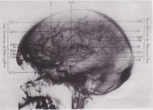
One of the first reports and image of cerebral angiography. From, Moniz E, de Carvalho L, Lima A. La radioartériographie et la topo-graphie cranioencephalique. J Radiol Electrol Med Nucl 1928;12:72. (Provided by Prof. Filipe Caseiro Alves)

At the beginning, Caseiro Alves plans to go on a short time travel through the history of Portuguese radiology. “Portugal’s greatest contributions to radiology are closely interwoven with the Portuguese School of Angiography, which began with the work of Egas Moniz, who was the first to perform a cerebral arteriography in 1927,” he explained.

Since then, Portugal has yielded many outstanding pioneers in the field of vascular radiology, among them Reynaldo dos Santos, a professor of surgery in Lisbon, who, among others, is known for the invention of aortography in 1929. Others include Lopo de Carvalho, who successfully introduced pulmonary angiography in 1931, Álvaro Rodrigues, Sousa Pereira and Roberto de Carvalho, who are known for their innovative work on lymphography (1933), Reynaldo’s son, João Cid dos Santos, who successfully introduced direct phlebography of the limbs in 1938, and Ayres de Sousa, whose name will always be closely associated with microangiography. “All these pioneer works were at the forefront of one of the most important achievements in healthcare and proved instrumental in shaping today’s medical practice,” Caseiro Alves said. Read more…

ECR continues to think outside the box and adds the Cube

For young physicians, interventional radiology (IR) may seem as enigmatic as a Rubik’s cube. But the cube can be solved with the right algorithm, according to Dr. Maximilian de Bucourt, head of angiography at Charité Campus Benjamin Franklin in Berlin.

“What we do in IR is exactly like a Rubik’s cube: you follow the algorithm and solve the problem. In IR, if you do the procedure steps over and over again and use the rules, most of the time you can get the solution for the patient,” he said.

Dr. Maximilian de Bucourt is head of angiography at Charité Campus Benjamin Franklin in Berlin, and one of the specialists behind the Cube.

Together with Prof. Christian Loewe from Vienna, de Bucourt imagined ‘the Cube’, a workshop aimed at introducing young physicians to the tools and techniques used in IR. With its focus on hands-on activities, including simulated procedures and interactive demonstrations, the Cube is fulfilling its goal of acquainting residents with this unique sub-specialty.

“The Cube is for young people who are thinking of becoming IR specialists, but find it too long before they can deploy a stent or manoeuver a catheter inside an artery. After medical school, basic radiology training and dedicated IR training with a teacher willing to let them perform major interventions, they still need to learn how to manage complications. The Cube is all about expediting this process, by enabling students and residents to get their hands on the products earlier,” he explained. Read more…

Riklund to unveil the next big thing in hybrid imaging

The Swedish radiologist Prof. Katrine Riklund, the current Chair of the ESR Board of Directors, has dedicated her career to the development of hybrid imaging. During todays’ Marie Curie Honorary Lecture (Room A, 1:00 p.m.) she will look back at the achievements made in this emerging field and look forward to future advances.

Riklund, who is a professor, consultant in diagnostic radiology and pro-vice-chancellor of Umeå University, is one of Sweden’s leading radiologists. She is also one of the first researchers to have recognised the potential of combining PET with CT and MRI, and has worked to advance the field ever since its emergence at the beginning of the 21st century.

“The combination of structural and functional/molecular imaging is fascinating. The entire field is new and extremely interesting, and it’s the closest to my heart when it comes to imaging,” she said.

Prof. Katrine Riklund from Umeå, Sweden, will speak about the growing significance of hybrid imaging in today’s honorary lecture.

During her lecture, Prof. Riklund will share examples of what hybrid imaging can do and what is going to come next. A major trend will be making use of the entire hybrid imaging examination for diagnostic protocols, also for the CT part, she believes. “This changes workflow and gives us more information. PET and CT or MR are not competing techniques, they are complementary,” she said.

Tracer development is key for PET but digital detectors will also represent a major step forward. Currently, the need for a cyclotron for production of radionuclides hinders substantial distribution of scanners outside large centres. “To make hybrid imaging really take off, we need other forms of tracer production. I would like to see the tracer production work like a coffee machine – with buttons to select tracer and radionuclide,” she said.

In her day-to-day work, Prof. Riklund is involved in various research projects, such as COBRA, a prospective multimodal imaging study of dopamine, brain structure and function, and cognition; (PEARL-PD), 18F-FE-PE2I PET/CT, a study of dopamine transporters in early Parkinson’s disease, RECTOPET (REctal Cancer Trial On PET-MR/CT); and Prostate Cancer – PSMA and Acetate in PET/CT and PET/MR. These projects reflect her three major interests in the field; movement disorders and cognition in central nervous system, and prostate and colorectal cancer.

Read more…