Swiss prepare to unveil connection between chocolate and radiology

by Viviene Raper

How to create hand-made chocolate masterpieces isn’t your usual radiology talk but, in tomorrow’s session, radiologists with a sweet tooth can learn all about truffle making and other confectionery arts from a leading Swiss chocolatier – interspersed with more conventional talks about radiology in Switzerland.

Andreas Trumpler, managing director marketing and group CMO at Läderach chocolatier suisse, will be giving two short presentations about how Swiss chocolate is transformed from cacao bean to chocolate, and then into a hand-made masterpiece. The company employs more than 800 people and since 1962, it has established a reputation for chocolate and confectionery specialties. Läderach’s products are sold throughout Switzerland and Germany, as well as by partners in the Middle East and Asia, and it supplies the top end of the gastronomy and hotel industry with semi-finished and finished products.

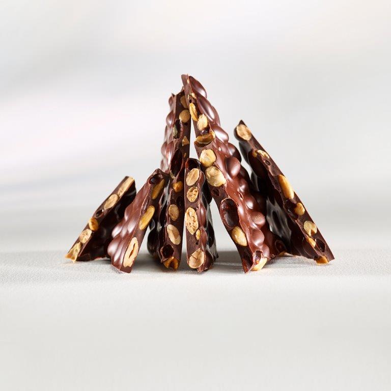
Fresh Chocolate with almonds from Läderach chocolatier suisse: the original view.

The connection between Swiss chocolate and Swiss radiology is one of quality, according to Prof. Dr. Dominik Weishaupt, who has been President of the Swiss Society of Radiology (SGR-SSR) since 2016 and is chairing tomorrow’s session.

“Over the 100 years of the Society’s history, we have undertaken several initiatives to improve the quality of Swiss radiology. And chocolate is considered as one of the highest quality products manufactured in Switzerland,” he noted.

Weishaupt, who is chief physician in the Institute of Radiology and Nuclear Medicine at City Hospital Zurich, explained that Swiss chocolate needs precision manufacturing, and Swiss radiology also prides itself on its attention to detail. He said that the SGR-SSR was the first medical society to introduce board exams, as well as clinical audits for radiology departments in the private and public sector, which they developed in close collaboration with the Ministry of Health. Read more…

The future is now, Marc Dewey says

The radiological community must understand the potential of value-based radiology and its related challenges, the German radiologist Marc Dewey will argue during the Wilhelm Conrad Röntgen honorary lecture today at ECR 2018.

Marc Dewey is Heisenberg Professor of Radiology of the German Research Foundation and Vice Chair of the Department of Radiology at Charité – Universitätsmedizin Berlin. His main research interest is in cardiovascular imaging but he is interested in many other aspects of clinical practice including value-based imaging, something he will detail in his lecture at the ECR.

Prof. Marc Dewey from Berlin will deliver today’s Honorary Lecture on value-based imaging at 12:15 in Room A.

“Participants will get to know three things. First, why the time for value-based radiology is now. Second, they will appreciate the potential of value-based radiology in the clinical care process. And third, understand the challenges in implementing value-based radiology,” Dewey promised.

Combining human image analysis and artificial intelligence has great potential for creating value for patients at lower costs, he believes. “This is value-based radiology and the time to get involved is now, as this is not merely a new technical toy of radiologists but will be accompanying the entire diagnostic and treatment pathway in all clinical service lines for the benefit of our patients,” he said.

Dewey is the coordinator of the DISCHARGE trial, an EU-funded multicentre project with more than 30 partners across Europe that will determine whether cardiac CT may replace invasive coronary angiography in certain patients. “The DISCHARGE project is a unique and truly impressive effort of several hundred individuals working at 31 sites in 18 European countries. External advisory board members were often thrilled when first witnessing the team spirit at our annual meetings,” said Dewey, who will soon present results of the on-going clinical trial. He said coordinating a large project on imaging in Europe gave him “the ability to better understand the culture, concerns, and ideas in different regions of Europe.”

Read more…

ECR 2018 celebrates the diversity of radiology

by Prof. Bernd Hamm, ESR President

It is my great pleasure to welcome you to ECR 2018!

The European Congress of Radiology has attendees from all over the world – from over 140 countries and many diverse cultures. It is a multi-professional meeting where international experts can shake hands with students, medical residents exchange ideas with physicists, and radiographers share their perspectives with industry representatives.

I chose ‘Diverse & United’ as our congress motto this year, as radiology is such a diverse specialty, covering a huge range of medical and scientific topics: from ever more refined diagnostic options to image-guided minimally invasive treatment options. Alongside our diversity, as radiologists and radiographers we should also stay united, which is in the interest of our specialty and our patients. This is what our congress is: something to offer for everyone, regardless of profession, cultural background or specialisation.

Bernd Hamm, ESR President

ESR President Bernd Hamm is professor of radiology and chairman of all three merged departments of radiology at the Charité, Humboldt-Universität zu Berlin and Freie Universität. He is also clinical director of the Charité Center, which includes radiology, neuroradiology, nuclear medicine and medical physics.

Being Congress President in 2015 gave me the unique opportunity for re-evaluation and to implement new things that I feel strongly about as well as fine-tune features that already existed. It was generally important to me to introduce new ideas in order for those of you who’ve even been coming to Vienna for decades, just like me, to get the chance to experience multiple innovations.

MyT3 is a new session format, adapting the ECR even more to these fast-moving times. 240 daring colleagues will present their scientific thesis in just three minutes! As if this wasn’t dramatic enough, we decided to hold these speedy sessions on the Sky High Stage which overlooks the city of Vienna, as only the sky is the limit for this new generation of radiology professionals.

Additionally, we created another new session format, ‘Coffee & Talk’, which is highly interactive with much more time for discussion than usual and in a relaxed atmosphere, with the possibility to also enjoy a coffee or other hot beverage. The interesting lectures on offer in these sessions call for an exciting exchange, bringing together different statements and opinions as well as Viennese coffee culture.

For the first time, the CUBE will open its doors to you: a theme park for interventional radiology (IR), designed for residents who haven’t specialised yet. Challenges, quizzes, training and much more will be focused on IR in emergencies plus other everyday topics, including the aorta, oncology, peripherals, and stroke. Without wanting to give away too much, I recommend paying the Cube a visit during lunchtime for ‘the main event’: the daily highlight involving experts in the arena, less challenging as well as more challenging interventions and much more. Come and be part of it!

Read more…

ECR 2018: uniting radiologists to show diversity of discipline

An exclusive interview with the incoming ESR President

ECR Today spoke with the incoming ESR President, Prof. Bernd Hamm, from Berlin, Germany, to learn about his ideas for next year’s congress.

ECR Today: You were already ECR Congress President in 2015. How did you come to be president again in 2018?

Bernd Hamm is professor of radiology and chairman of all three merged departments of radiology at the Charité, Humboldt-Universität zu Berlin and Freie Universität (Campus Mitte, Campus Virchow-Klinikum, and Campus Benjamin Franklin). He is also clinical director of the Charité Center, which includes radiology, neuroradiology, nuclear medicine and medical physics.

Bernd Hamm: This is not the first time that I have been asked: “why are you president again?” It was a great pleasure and responsibility to be the ECR President in 2015. For many years, we had two presidents – one to manage the affairs of the European Society of Radiology (ESR) and another to organise the society’s annual congress (ECR). And frankly, in my opinion this was a good division of labour. However, “the times they are a changin’ …” and, therefore, our society changed its statutes, and now we only have one president, who is both President of the ECR and President of the ESR.

This new combined ESR/ECR President begins his or her term of office at the end of each ECR by organising and chairing the next ECR and then the following year, as Chairman of the Board, focuses on professional and administrative business of the ESR. For me, this means a double honour and double responsibility. When I was elected to be the congress president for ECR 2015, I made a great effort to make the ECR a success and I will do my best to make ECR 2018 a success too.

However, I wish to underline that the ECR is a team effort and that the Programme Planning Committee with its brilliant members is a major asset in compiling an attractive scientific programme and that the organisation of a congress like the ECR is not possible without the highly professional staff of the ESR Office. Working together with many colleagues from many countries and different fields of radiology is very inspiring and is what ultimately underlies an attractive congress. This joint effort is rewarding and enlightening.

ECRT: As a motto for ECR 2018 you chose ‘Diverse and United’. Could you tell us a little about this?

BH: Radiology is such a diverse specialty, ranging from more and more refined diagnostic options to image-guided minimally invasive treatment options. Our specialty has something to offer for all of us and also for future generations of physicians, radiographers and students. However, like in a mosaic, it is the connection of all the pieces that create the big picture. By using this metaphor I wish to express my sincere wish and hope that, despite these different and interesting facets and subspecialties, radiologists should see themselves as a community and join forces to strengthen our specialty in the best interest of our patients.

In 2015 we also had a motto – ‘Radiology without borders’. In the beginning of 2015, none of us would have expected to see new fences being erected in Europe and borders becoming important once again. In view of these tendencies, it is even more important for our scientific community to practice radiology without borders and to advance our specialty through free academic discourse and by benefiting from a diversity of ideas.

Read more…

05
Mar 2017
POSTED BY
POSTED IN ,
DISCUSSION 0 Comments

European Radiology: 25 years of excellence

Watch the European Radiology 25th Anniversary session live on ECR Online, Friday, March 3, 16:00–17:30, Room Z
#ECR2017 

Albert L. Baert, Adrian K. Dixon, Maximilian F. Reiser.

25 years of success for the three editors-in-chief of European Radiology: Albert L. Baert, Adrian K. Dixon, Maximilian F. Reiser.

The ESR’s flagship journal, European Radiology, celebrated 25 years of publication in 2016. We spoke to Prof. Albert L. Baert, the journal’s Editor-in-Chief from 1995 to 2007, Prof. Adrian K. Dixon, who headed European Radiology from 2008 until 2013, and our current Editor-in-Chief, Prof. Maximilian F. Reiser. Together, they gave us a comprehensive account of the journal’s development during the last quarter of a century.

 ECR Today: How have you seen the journal develop during the past 25 years?

Albert L. Baert: During this time period the number of published articles has increased spectacularly from 60 articles per year in the first issues to more than 400 per year now. For example, no less than 4,674 pages were published in 2016! European Radiology is now one of the most widely distributed and read scientific journals in the world. Simultaneously, the scientific level of the contents has improved steadily over the years as proven by the current excellent Impact Factor ranking.

Read more…

02
Mar 2017
POSTED BY
POSTED IN ,
DISCUSSION 0 Comments

Facial genetics and forensics take centre stage at ECR

Watch this session on ECR Live: Friday, March 3, 10:30–12:00, Room B
#ECR2017

Forensics and facial genetics will be in the spotlight at ECR 2017 as the ‘ESR meets Belgium’ session offer a look at medical imaging’s most original contributions to healthcare and crime investigation.

Advanced decomposition of brain MRI or facial 3D surface images into modules, tailored for associations with underlying genetic variations.

Dr. Peter Claes from Leuven, Belgium, will present his work in facial genetics in a session titled ‘Imaging genetics and beyond: facial reconstruction and identification’.

Claes is a senior research expert in the Medical Image Computing research group of the Processing of Speech and Images division of the Electrical Engineering department at Leuven Catholic University.

He uses CT, MRI and 3D surface imaging modalities to grasp the link between people’s appearance and underlying genetic variations.

“Your appearance is genetically driven. In families there’s a strong link, even more so between identical twins, who share the same DNA profile and almost the same face. Physical features also influence your brain, the way you think. A lot of facial characteristics are shared, for example in Down’s syndrome patients, who present with the same features whether they are European or Asian,” he explained.

The link between genetic disorders and facial genes has been of interest to scientists for a while, but research is slow and tedious.

Claes became interested in facial genetics after working in craniofacial morphometrics to help correct morphological abnormalities and anomalies, and in craniofacial reconstruction to identify victims.

To help decipher facial genetics, Claes uses the computer-based craniofacial reconstruction programme he developed for victim identification, and combines 3D surface processing, statistical modelling, analysis, mapping and prediction techniques. He has also created an array of algorithms and software for investigators who plan to use 3D facial datasets. Last year, he also co-organised the first international workshop on facial genetics in London.

Read more…

Celebrating Ten Years of ESOR

Guest article by ESOR Director, Prof. Nicholas Gourtsoyiannis

Nicholas Gourtsoyiannis

Nicholas Gourtsoyiannis is the Educational & Scientific Director of the European School of Radiology (ESOR) and chairman of the ESR’s ESOR Committee.

The European School of Radiology (ESOR) has completed ten years in action. Ten challenging and rewarding years of unfailing commitment and continuous investment in radiological education in Europe. Ten years of envisioning, engaging, delivering, teaching, tutoring, nurturing, and adding value to radiology.

The three main goals of ESOR are still to assist in harmonising radiological education throughout Europe, by supporting the implementation of the European Training Curricula (ETC); to build a genuine and firm interest in subspecialisation in radiology; and to raise the scientific profile of radiological education in Europe and worldwide.

The past ten years of ESOR have been marked by an outstanding growth in a wide range of modular activities, including foundation and advanced courses, teach-the-teachers and visiting professorship programmes, visiting schools, seminars, tutorials, preparatory courses for the European Diploma in Radiology (EDiR), scholarships, exchange programmes for fellowships and full one-year fellowships. So far, ESOR has delivered structured continuing education to almost 17,000 residents and board-certified radiologists worldwide, through 800 programmes.

Read more…

02
Mar 2017
POSTED BY
POSTED IN ,
DISCUSSION 0 Comments

Education central to improving imaging data quality in oncology clinical trials

Watch this session on ECR Live: Thursday, March 2, 16:00–17:30, Room X
#ECR2017

Imaging data is key in multicentre clinical trials for cancer research but quality control is currently a major impediment, bringing the validity of the trials into question and potentially impacting on the quality of drugs put on the market, a panel of experts will argue today in a session held by the ESR and the European Organisation for Research and Treatment of Cancer (EORTC) at the ECR.

Imaging is increasingly contributing to cancer research thanks to the development of innovative techniques that depict functional and molecular processes. In most oncological clinical trials, imaging is now the primary criteria used to evaluate progression of disease or efficiency of the drug being tested.

The best way to obtain valuable imaging measurements is to involve the imagers who take part in these trials and educate the clinician investigators, experts will explain in the session.

When it comes to imaging in cancer research, a number of issues take centre stage. Difficulties associated with integrating imaging biomarkers into trials have been neglected compared with those relating to the inclusion of tissue and blood biomarkers, largely because of the complexity of imaging technologies, safety issues related to new contrast media, standardisation of image acquisition across multivendor platforms and various post-processing options available with advanced software, as reported recently in The Lancet by the EORTC and leading researchers.

Read more…

01
Mar 2017
POSTED BY
POSTED IN ,
DISCUSSION 0 Comments

Eminent neuroradiologist to give honorary lecture at ECR 2017

Watch this session on ECR Live: Thursday, March 2, 12:15–12:45, Room A
#ECR2017

In recognition of his significant impact on the field of neuroimaging and his service to organised radiology, Professor Mauricio Castillo from Chapel Hill, NC, United States, has been invited to deliver the Wilhelm Conrad Röntgen Honorary Lecture ‘Dissatisfaction, burnout and inequality: three major challenges in radiology’ at ECR 2017.

Professor Mauricio Castillo from Chapel Hill, United States

Professor Mauricio Castillo from Chapel Hill, United States, will deliver the Wilhelm Conrad Röntgen Honorary Lecture today at 12:15 in Room A.

Mauricio Castillo is the James H. Scatliff distinguished professor of radiology, chief and programme director of neuroradiology at the University of North Carolina in Chapel Hill. He is currently president of the American Roentgen Ray Society.

Originally from Guatemala, Prof. Castillo completed his radiology and neuroradiology training at the University of Miami School of Medicine, Jackson Memorial Medical Center, and Emory University School of Medicine, Affiliated Hospitals in Atlanta respectively.

His specialty and subspecialty choice was in large part motivated by his mentors.

“When I was a medical student, I was fortunate to work in the only teaching hospital that had a CT scanner and two board-certified radiologists. I was very impressed by the impact of imaging in patient care and by the way these two professionals used their large base of knowledge of medicine to generate a differential diagnosis based on imaging findings. Later on, while I was a resident, I developed a close relationship with Dr. Robert Quencer, which led me to become a neuroradiologist. One should never underestimate the influence we may have on our trainees and the influence our mentors have had on us,” he said.

Prof. Castillo’s research interests include paediatric neuroimaging, application of new imaging techniques and medical literature editing. Read more…

Image-guided interventions: a key pillar in cancer care

Watch this session on ECR Live: Wednesday, March 1, 16:00–17:30, Room F2
#ECR2017

 There is hardly any area of hospital medicine where interventional radiology (IR) has not had some impact on patient management. The range of conditions that can be treated using interventional radiology techniques is continually expanding.

In today’s session, experts will provide an insight into image-guided interventions in oncology with a particular focus on illustrating the importance of quality assurance in image-guided oncological interventions and their effect on treatment outcomes.

In recent years, IR has played a vital role in the field of oncology, and alongside medical, surgical and radiation oncology it constitutes a key pillar in cancer care. Vascular and non-vascular procedures such as transarterial chemoembolisation, radiofrequency ablation (RFA), microwave ablation, radioembolisation, cryoablation and high-intensity focused ultrasound (HIFU) are delivered locally, minimise damage to nearby tissue and avoid the systemic side effects of chemotherapy.

Colorectal lung metastasis before CT – guided microwave ablation.

For the interventional radiologist providing oncologic therapies it is essential to understand the rapidly changing field of oncology and to have a broad knowledge of oncologic diseases and available therapies to treat them. Radiologists providing image-guided interventions in oncology have an outstanding understanding of imaging as well as a diversity of interventional skills. However, they lack formal training in oncology and an understanding of chemotherapy and radiotherapy, according to Prof. Andy Adam from the Department of Radiology at Guy’s and St. Thomas’ Hospital in London. Read more…