Prominent Brazilian radiologist to shed light on Zika virus imaging at ECR 2017

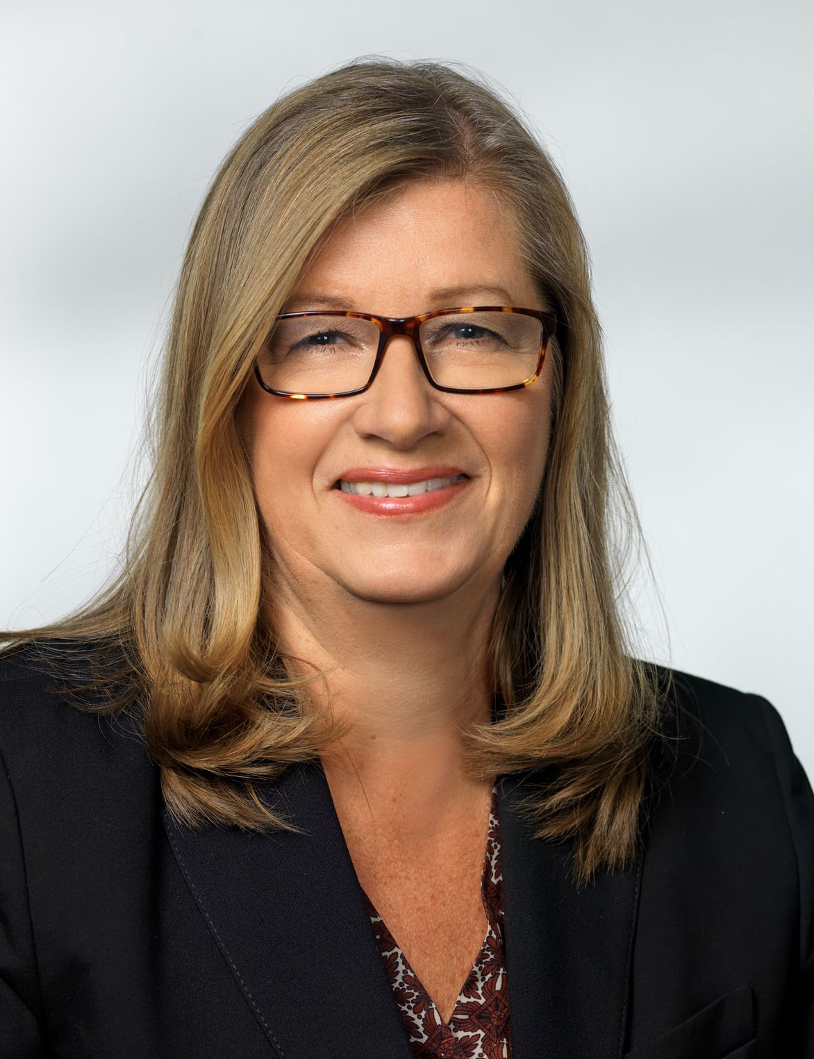
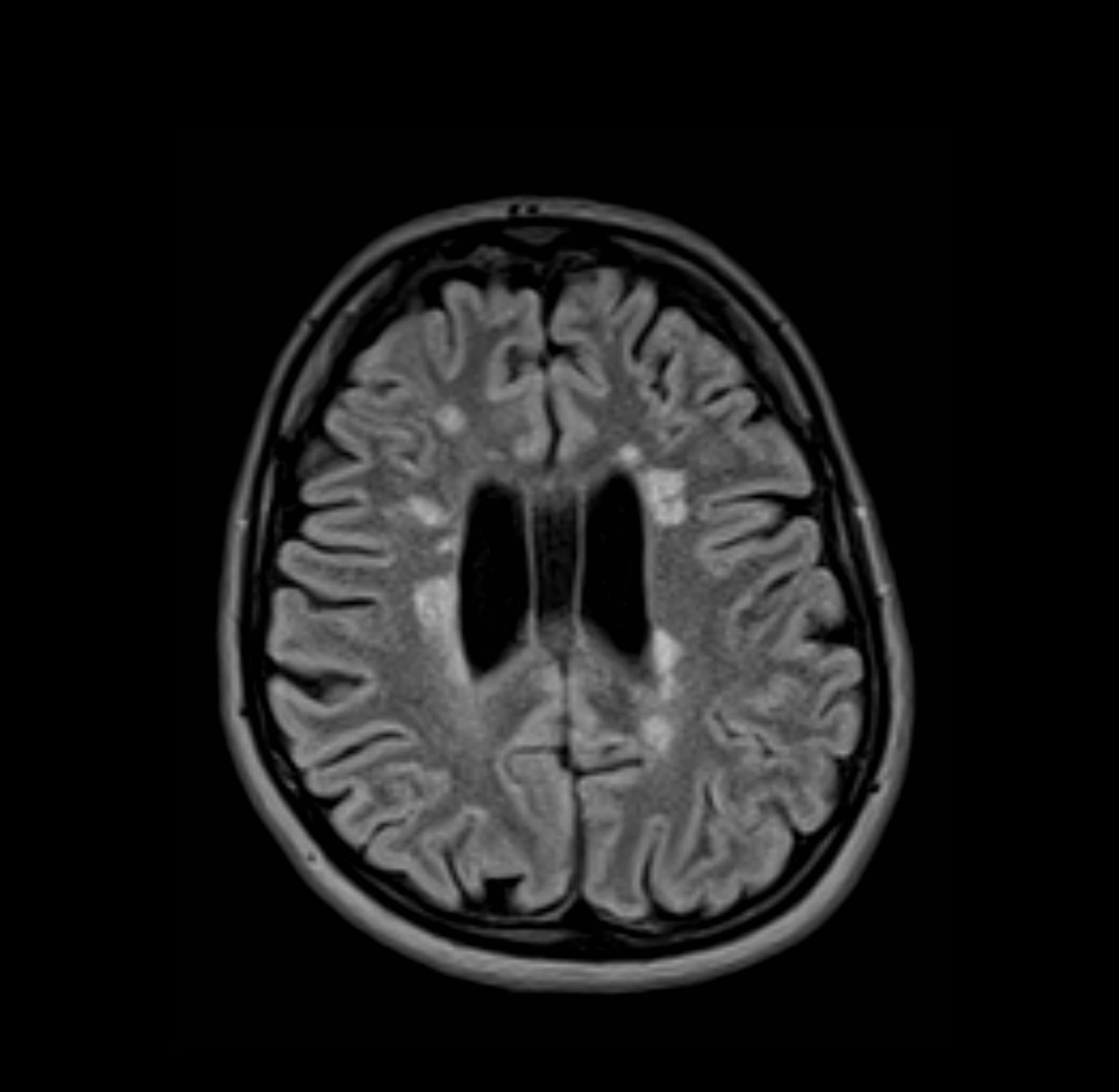
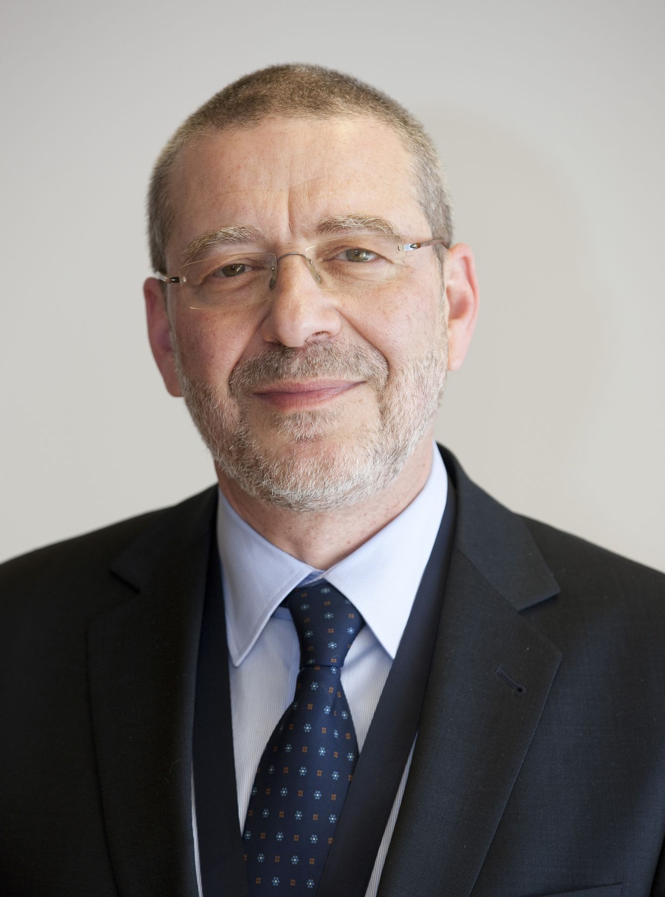
In recognition of her major achievements in neuroimaging and advancement of the field, Professor Maria de Fátima Viana Vasco Aragão from Recife, Pernambuco, Brazil, will present the Guest Lecture ‘Breaking News from Latin America: How to recognise Zika virus infections on imaging studies’ at ECR 2017.

Maria de Fátima Viana Vasco Aragão is professor of radiology at the Maurício de Nassau University and Scientific Director of the Multimagem Diagnostic Centre in Recife, Brazil. She is also financial director of the Diagnostika Endoscopy and Radiology Clinic in Recife.

Prof. Vasco Aragão received her medical degree from Pernambuco Federal University in 1987 and completed her residency in radiology at Ribeirão Preto Faculty of Medicine, University of São Paulo. She did fellowships in CT and MRI and in neuroradiology at the Med Imagem Beneficencia Portuguesa, São Paulo, and later completed a research fellowship at Mount Sinai Hospital Center in New York, US.

She always knew she would become a doctor and chose radiology because she recognised early on the field’s value in diagnosis and treatment.

Maria de Fátima Viana Vasco Aragão is professor of radiology at the Maurício de Nassau University and Scientific Director of the Multimagem Diagnostic Centre in Recife, Brazil.

Maria de Fátima Viana Vasco Aragão is professor of radiology at the Maurício de Nassau University and Scientific Director of the Multimagem Diagnostic Centre in Recife, Brazil.

“My mother is a retired biologist and biology teacher. I have never forgotten the moment she taught me what a cell was. In my family, educators predominated and my great aunt was the first woman in Pernambuco, my home state, to graduate from medical school, in 1935. Perhaps on account of this family background I made up my mind to become a doctor. As an intern student of medicine, I was very impressed when I took my patients to be examined by CT and US. Even though there was only one CT scanner in my city, I succeeded in having all my intern patients examined, when indicated. My tutors were impressed by the swiftness with which cases were resolved. Thus I learned that radiology not only helps to quickly define diagnosis in most patients, but also provides a clear prognosis for the most adequate treatment. At the end of the course I began considering the possibility of specialising in radiology, even though 29 years ago, for most people, a radiologist was not a doctor. Shortly after graduation, I sat for a test and won a place in radiology at the first attempt, and radiology has been my passion ever since. I would like to say that I am grateful for having had great mentors, including the neuroradiologist Dr. Sérgio Santos Lima, who was my director during my fellowship in the Med Imagem Beneficencia Portuguesa, and the head and neck radiologist, Dr. Peter Som, who was one of my directors during my research fellowship at Mount Sinai Hospital Center New York,” she said.

Read more…

New ESR/ECR president to make youth a central theme at ECR 2017

ECR2016_ECRToday_Blog

ECR Today spoke with the new ESR/ECR President, Prof. Paul M. Parizel, from Antwerp, Belgium, to learn about this new position, his visions for the society and his ideas for next year’s congress.

Prof. Paul M. Parizel, chairman of Antwerp University Hospital’s department of radiology and full professor of radiology at the University of Antwerp’s faculty of medicine, is the incoming ESR/ECR President.

Prof. Paul M. Parizel, chairman of Antwerp University Hospital’s department of radiology and full professor of radiology at the University of Antwerp’s faculty of medicine, is the incoming ESR/ECR President.

ECR Today: You are the first officer of the European Society of Radiology to take on the new position of combined ESR/ECR President. Could you please briefly explain to our readers how this change came about and what it means? What are your main tasks and responsibilities in this position?
Paul M. Parizel: It is a great honour, and also a huge responsibility, to assume this new position of combined ESR/ECR President. The ESR is one of the most important and prestigious international scientific societies, with more than 63,000 members throughout the world. The ECR is well established as the foremost congress in radiology in Europe. Until a few years ago, nominations and elections for the ‘cursus honorum’ of the ECR and ESR were made independently of each other. This implies that we have had brilliant presidents of our society, who never became president of the congress, and vice versa. As both the society and the congress were getting bigger, and more mature, it was decided to re-evaluate and retune the strategic plan and to change the statutes so that appointments were fully integrated, instead of running on parallel tracks, as was previously the case. Bringing the congress and the society under one and the same leadership umbrella is an efficient way to better utilise our resources (human, political and financial). I am convinced that this will improve communication with our members, enhance our international standing and facilitate relationships with other societies and with the industry.
This combined ESR/ECR presidency is certainly a daunting task, and I admit that I am a little bit nervous. On the other hand, I can rely on the work of my predecessors and especially on the creative input and unflagging energy of my friends and colleagues of the Board of Directors, the Executive Council, the statutory committees, subcommittees, working groups, and, last but not least, the experience, professionalism and efficiency of the ESR staff. Our society is healthy, both politically and financially, and we are steering a stable and steady course.

Read more…

Hybrid imaging contents to heighten delegates’ interest at ECR 2016

ECR2015_ECRToday_Blog

ECR Today spoke with ECR 2016 Congress President, Prof. Katrine Åhlström Riklund, deputy head of the department of radiation sciences and director of the medical school at Umeå University, Sweden, to find out a little bit about next year’s annual meeting.

ECR 2016 Congress President, Prof. Katrine Åhlström Riklund, from Umeå, Sweden.

ECR 2016 Congress President, Prof. Katrine Åhlström Riklund, from Umeå, Sweden.

ECR Today: What will be the highlights of ECR 2016?
Katrine Åhlström Riklund: It is hard to tell what the specific highlights will be more than one year ahead, due to the rapid development of imaging. The highlights will be the entire congress through its well-developed programme, which covers the whole range of education from student level to advanced subspecialists. I should say the added content of hybrid imaging in several sessions would make the programme even more attractive. Besides the educational and scientific programme, the grand opening ceremony and social activities will also be memorable events.

ECRT: Will there be any new additions to the programme?
KAR: As always, there will be innovations at the ECR. The content of hybrid imaging will be spread across several sessions and not in one single session. The new session formats introduced at ECR 2015, with the European Excellence in Education (E3) programme – divided into five levels (the Rising Stars programme, European Diploma Prep Sessions and Beauty of Basic Knowledge programme, ECR Academies and ECR Master Classes) will be continued. These levels cover the entire span from undergraduate medical education to subspecialised continuing professional development. Getting involved in the sessions is important for retaining knowledge.

Read more…

ESR to address issues on all fronts in 2015

ECR2015_ECRToday_Blog

ECR Today spoke with the incoming ESR President, Prof. Lluís Donoso Bach, Director of the Diagnostic Imaging Department of the Hospital Clínic of the University of Barcelona and Executive Director of the UDIAT Diagnostic Centre at the Health Corporation Parc Taulí, to find out about his plans for the ESR over the coming year.

New ESR President, Prof. Lluís Donoso Bach

New ESR President, Prof. Lluís Donoso Bach

ECR Today: What will be your priorities as ESR President?
Lluís Donoso Bach: The ESR has a good track record in education and we will continue to innovate through the ECR. We will also open new learning centres in Vienna and Bogotá and are planning a further centre in Moscow.
Because of the financial crisis, it is more important than ever that we develop efficient systems, so we will increasingly rely on electronic, web-based services for our members. This is why we decided to launch the ESR eLearning Platform. This represents a big challenge, and we will see how it works as a business model and how our members use the platform throughout the year. We will also try to offer an examination for the European Diploma in Radiology online. There are a lot of experiences and successful models to draw inspiration from.
We will also continue our efforts in research through the European Institute for Biomedical Imaging Research (EIBIR). The ESR Research Committee has been working a lot on the quantification of data using biomarkers and biobanks, focusing in particular on oncology and trying to have more influence in that area. Quality and safety is another very important issue for us. During ECR 2015, we launched ESR iGuide, a clinical decision support system for European imaging referral guidelines, following the example set by the American College of Radiology with ACR Select.
We also want to offer our members tools to improve quality in their departments. The ESR Quality, Safety and Standards Committee has thus created Level 1 clinical audit templates based on safety. We should also explore the possibility of performing quality controls at the level of department management.
Lobbying with European institutions has been an important field of battle for us in the past few years, and we will consolidate our action in this field keeping the momentum and relying on the personal contacts and networks established over the past years. We launched a Call for a European Action Plan for Medical Imaging in the European Parliament on November 4, to highlight existing heterogeneities in medical imaging in Europe in the areas of quality and safety, education and training, research, and eHealth, and to call for joint targeted actions to improve harmonisation in these areas.

Read more…

08
Mar 2015
POSTED BY
POSTED IN , ,
DISCUSSION 0 Comments

Comprehensive personalised imaging transforms cardiothoracic disease management

ECR2015_ECRToday_Blog

Watch this session on ECR Live: Sunday, March 8, 8:30–10:00, Room E1
Tweet #ECR2015E1 #NH17

Besides personalised imaging, a new paradigm is emerging in radiology that should re-shape clinical practice and benefit the patient immensely. Supported by new technologies that enable radiologists to image the body faster and better, radiologists are now trying to broaden their focus during examinations.

If there is a field where these advances make a tremendous difference, it is cardiothoracic imaging, an area where diseases are more often than not intertwined. Cardiovascular and chest radiologists will explain how the comprehensive personalised approach impacts their work and try to convince radiologists on both sides to take an interest in the other, in a New Horizons session on Sunday at the ECR.

For years, the trend was for radiologists to subspecialise as much as they could. Cardiovascular radiologists and chest radiologists would focus on their own area with little or no interest beyond that. But among these subspecialists, an increasing number are now changing their approach, as mounting evidence shows that diseases of the heart and chest are very often related, according to Dr. Christian Loewe, deputy head of the section of cardiovascular and interventional radiology at the Medical University of Vienna, Austria.

Dr. Christian Loewe is deputy head of the cardiovascular and interventional radiology section at the Medical University of Vienna, Austria.

Dr. Christian Loewe is deputy head of the cardiovascular and interventional radiology section at the Medical University of Vienna, Austria.

“In the past patients were investigated by either focusing on chest or cardiac diseases. This choice was mainly driven by their first clinical examination. However, there are a lot of situations and diseases where chest problems are caused by cardiac diseases and vice versa. There’s a huge interaction between heart and chest, and that’s why it’s interesting and important to look at this relationship in more detail today,” he said.

To prove his point, Loewe, a cardiovascular radiologist, will talk about acute and chronic chest pain during the session. Some of the most severe causes of chest pain are due to cardiovascular diseases, including myocardial infarction or acute aortic diseases. However, acute chest pain can also be caused by a number of pulmonary diseases, including pulmonary embolism, pneumonia and others. Therefore, radiologists must learn the different life-threatening disorders that cause chest pain, whether they are respiratory or cardiovascular.

Read more…

New opportunities for partnership between radiology and radiation oncology

ECR2015_ECRToday_Blog

Since the discovery of the x-ray, radiology and radiation oncology have been sister disciplines. Recent progress has brought increasing points of interaction between the two and this is no better exemplified than by the close relationship between the ESR and the European Society for Radiotherapy and Oncology (ESTRO). The two societies will host a joint session at ECR 2015 focusing on this partnership and opportunities for future collaboration. To find out more about the session, ECR Today spoke to session co-chairman and ESTRO president, Prof. Philip Poortmans, of the Radboud University Medical Center, Nijmegen, Netherlands.

Prof. Philip Poortmans, President of the European Society for Radiotherapy and Oncology, will moderate today’s joint session on radiology and radiation oncology.

Prof. Philip Poortmans, President of the European Society for Radiotherapy and Oncology, will moderate today’s joint session on radiology and radiation oncology.

ECR Today: How was this joint session between the ESR and ESTRO first initiated?
Philip Poortmans: The time of medical specialties working on their own is over. It is now all about cooperation and multidisciplinarity. Often when we talk about multidisciplinarity, people think only about doctors who are directly involved in treating the patient. In the case of breast cancer, for example, this would then be the surgeon, the radiation oncologist, the medical oncologist and in several countries also the gynaecologist. People often forget about diagnostic specialists like the pathologist and the radiologist. Radiation oncology is very closely linked to imaging in general; both to radiology and to nuclear medicine. So it is a field that is very important for us and a specialty with which it is essential for us to cooperate closely. For many years, ESTRO has run courses with contributions from radiologists and nuclear medicine specialists to teach our young colleagues, or colleagues who want CME, about the contribution of the diagnostic specialties. So this is not new at all. ESTRO and the ESR have an especially close relationship, with the former president of ESTRO, Prof. Vincenzo Valentini, and the ESR president, Prof. Lorenzo Bonomo, working in the same hospital. People in such positions who know each other very well can facilitate this process of close collaboration which then benefits us all. Of course, this means that last year we already had a joint session at the ECR. This collaboration is based on a Memorandum of Understanding signed by both societies, which includes agreements about education, guidelines, and scientific dissemination. A congress is of course always a mixture of both scientific dissemination, bringing new findings to the community, and education, so this nicely fits our mutual commitment.

Read more…

Population imaging studies gain ground in healthcare

ECR2015_ECRToday_Blog

Watch this session on ECR Live: Thursday, March 5, 16:00–17:30, Room L1
Tweet #ECR2015L1 #PC8B

Imaging large cohorts of people enables scientists to collect information useful for science and emphasises radiology’s role in healthcare. From the most recently available imaging biomarkers to data such as genomics and metabolomics, Thursday’s dedicated Professional Challenges Session will show just how useful population imaging studies have become in the prognosis of countless diseases.

When radiologists follow a cohort of people for 20 or 30 years, the benefits for the patient increase tremendously. If a radiologist performs a CT examination of a patient’s coronary arteries and finds calcification, chances are that the patient will have a heart attack within the next few years. Unfortunately at this stage, the patient is usually out of the radiologist’s reach.

The fluid-attenuated inversion recovery axial image of a 26-year-old female participant shows multiple, bilateral, asymmetric, linear and ovoid hyperintensities that are located perpendicular callososeptal characteristic of multiple sclerosis

The fluid-attenuated inversion recovery axial image of a 26-year-old female participant shows multiple, bilateral, asymmetric, linear and ovoid hyperintensities that are located perpendicular callososeptal characteristic of multiple sclerosis

However, if patients chose to participate in a population study, they will be checked on a regular basis, and radiologists will be able to access previous information and initiate appropriate treatment earlier, significantly
improving patient outcome.

Securing imaging data is always tricky and population imaging studies are an opportunity for radiologists to access this data. Showing the relevance of imaging findings highlights radiology’s role in the medical continuum, according to Prof. Norbert Hosten, of the Ernst-Moritz-Arndt University in Greifswald, Germany, who will chair
the session Thursday.

“Our way to prove that radiology can make people healthier and happier is to do large population imaging studies. Radiology can develop the kind of data that are necessary to prove that our methods really help the patient,” he said.

Read more…

Experts look into radiology’s future

ECR2015_ECRToday_Blog

Watch this session on ECR Live: Thursday, March 5, 8:00–10:00, Room E2
Tweet #ECR2015E2 #PC5A

Radiology is changing. Its possibilities are expanding; its place in healthcare has evolved. However, it has also become more vulnerable to financial turmoil. The future of the profession will depend on how radiologists decide to act and how well they can cope with external factors, a panel of experts will explain during today’s Professional Challenges Session at the ECR.

Prof. Gabriel Krestin, from Erasmus MC, University Medical Center Rotterdam, will speak about upcoming challenges for radiologists in today’s session.

Prof. Gabriel Krestin,
from Erasmus MC,
University Medical Center
Rotterdam, will
speak about upcoming challenges
for radiologists in today’s session.

The emergence of quantitative imaging and the development of imaging biomarkers are transforming the face of radiology. An increasing number of biomarkers are being validated and accepted as measures for prognosis, diagnosis or therapy monitoring. The transition from research to clinical practice has started in many areas, and clinical radiologists have become aware of this evolution. They must learn how to use these new tools, but it is tempting to resist change, especially when it brings extra work, according to Prof. Gabriel Krestin, professor and chairman of the department of radiology at Erasmus MC, University Medical Center Rotterdam, the Netherlands.

“Radiologists don’t like to perform measurements in daily practice because they are not used to it. They haven’t been trained for that and it’s time consuming. For instance, if you do a cardiac examination, extracting quantitative data from that examination takes up to 30 minutes, so you need dedicated personnel to do that – preferably radiographers. The workflow of the radiologist could be seriously disrupted if he/she had to perform such extensive post-processing of images, and the consequence would be a decrease in efficiency, particularly because nobody pays for the additional measurements,” he said.

Read more…

04
Mar 2015
POSTED BY
POSTED IN ,
DISCUSSION 1 Comment

Experts sound the alarm on dangers facing radiologists

ECR2015_ECRToday_Blog

Watch this session on ECR Live: Wednesday, March 4, 16:00–17:30, Room E2
Tweet #ECR2015E2 #PC4A

Remember HAL 9000, the murderous computer in ‘2001: A Space Odyssey’? This scenario doesn’t seem too far off when it comes to radiologists and the constant evolution of their technology. Luckily, they can still stay in control if they change their behaviour and remain at the head of the imaging process, experts will argue during a Professional Challenges session today at the ECR.

Radiologists must claim their place in the medical team if they are to survive. They must come out of the shadows and show that they can bring added value to the team, according to Jim Reekers, professor of interventional radiology at the University of Amsterdam, who will chair the session.

Jim Reekers, from the University of Amsterdam, will chair the session on the role of the radiologist.

Jim Reekers, from the
University of Amsterdam, will
chair the session on the role of the
radiologist.

“I want to make my colleagues aware that they should step out of their offices and act as doctors who work in multidisciplinary teams. Being the photographers or doctors who only give a report with a picture will not secure their future and the reason is very simple: anybody can make and look at a picture nowadays. The difference the radiologist brings is that he or she can give an interpretation of the picture within the context of a patient’s clinical situation. So the radiologist should become a doctor who gets involved with the patient’s situation,” he said.

There is more than one way to do this. For starters, to be on the same level as clinicians, radiologists must behave as such. That means they have to do more than just show the images in multidisciplinary team meetings, they have to sit and discuss them with the doctors.

“Have a junior show the images for you. While my junior presents the pictures, I am sitting together with the other specialists, and suggesting treatment and follow-up options or further diagnostic investigation. It is important to sit with the other doctors. There’s a big difference in how people see me. I’m one of them instead of being the guy who shows the pictures,” Reekers said.

Read more…

New treatments give hope to hearing impaired

ECR2015_ECRToday_Blog

Watch this session on ECR Live: Wednesday, March 4, 08:30–10:00, Room E1
Tweet #ECR2015E1 #SF1B

Hearing loss can present many difficulties and obstacles to sufferers, and with ageing populations it’s set to become a major healthcare challenge. Many conditions such as congenital malformation of the inner ear or hypoplastic cochlear nerve can also lead to hearing loss, and sometimes deafness.

Fortunately, many new treatments are available to recover hearing, both partially and completely. Imaging plays an increasingly important role in therapy planning and follow-up, and there is hope on the research front, experts will show during a dedicated Special Focus session on Wednesday morning.

Microtia – congenital anomaly of external and middle ear, resulting in conductive hearing loss. External auditory canal is not patent (arrow), mastoid process is underdeveloped (arrowhead)

Microtia – congenital anomaly of external and middle ear, resulting in
conductive hearing loss. External auditory canal is not patent (arrow),
mastoid process is underdeveloped (arrowhead)

The prevalence of auditory problems in the Western world has doubled over the past 30 years. It is estimated that between 15 and 17% of the population will suffer hearing loss, due to ageing or congenital malformation, but also bad habits, according to Agnieszka Trojanowska, a radiologist at Lublin University Medical School, Poland, who will
chair the session.

“We start to see young adults in their early 30s with sensorineural hearing loss or other related problems because of high frequency noise, which is typical for listening to music. Twenty years ago, such a condition was linked with working in fabrics or on the street. But the good news is that even if you use your iPod a lot, the degree of hearing
loss is light to moderate, so this is not something that will considerably affect your life,” she said.

Read more…

03
Mar 2015
POSTED BY
POSTED IN ,
DISCUSSION 1 Comment