ECR 2013 Rec: Ultrasound elastography #A028 #RC302

ECR2013Rec_Blog_Cover_RC302_A028

A-028 Ultrasound elastography

A. Athanasiou | Thursday, March 7, 16:00 – 17:30 / Room F2

Breast ultrasound elastography provides information about tissue elasticity Young modulus E = s / e, where s is the compression (stress) and e is the deformation (strain) of the tissue. It is a complementary tool to breast ultrasonography, easily performed in clinical practice. Two elasticity modes are currently available: strain imaging, where manual compression is applied to the ultrasound probe and tissue displacement is registered; tissue deformation is then calculated by means of dedicated software providing real-time elasticity images (color- or grey-coded) superimposed on B-mode imaging. This is a qualitative or semi-quantitative mode. Shear wave imaging, where US probe is used to induce mechanical vibrations using acoustic radiation force generating local tissue displacement. This mode provides quantitative information about either tissue displacement velocity or tissue stiffness itself in kPa. Functional information provided by elasticity imaging can be particularly useful for BIRADS 3 or 4a lesions. Various studies indicate that elasticity combined to B-mode imaging can improve breast ultrasound specificity up to 75-88%. False negative findings may be encountered in case of “soft” lesions (mucinous, medullary or cystic carcinomas) or inflammatory cancers. Differentiation between echogenic cysts and homogeneous solid lesions (such as fibroadenomas) can be improved as cystic features are usually specific in elasticity imaging. Iso-echoic lesions such as infiltrating lobular carcinomas may be better delimitated. Lymph-node characterization and microcalcification assessment can be improved, although few data are available and need further validation. 3D elastography is actually in progress and would be useful in monitoring response to neoadjuvant treatment.

17
Jul 2013
POSTED BY
DISCUSSION 0 Comments

ECR 2013 Rec: Finding the time and resources in the radiology department #A024 #PC3

ECR2013Rec_Blog_Cover_PC3_A024

A-024 Finding the time and resources in the radiology department

J. del Cura | Thursday, March 7, 16:00 – 17:30 | Room F1

One of the problems of undergraduate teaching of Radiology is the lack of time for teaching, due to competition for resources with other academic disciplines. Available classroom time and hours of practice are often insufficient to teach the increasingly complex modern Radiology. Also, the availability of financial resources to hire staff or access to educational facilities is competitive and limited, especially in a context of economic crisis. A good solution is to shift the paradigm of education, changing theoretical teaching into self-learning by students. This change allows to free class time to effectively teach Radiology. Classes are converted in workshops, doubt-solving sessions and problem-based learning, all of which matches better with a visual discipline like Radiology. Both on-line classes and e-learning can be useful for this purpose. This kind of teaching also makes Radiology a very attractive discipline for Medicine students. Also, Radiology practices can be carried out using custom computer applications. The lack of professors (and time) for practices can be solved with the help of residents, who are willing to participate as they are more prone to understand the learning needs of the students. Finally, the lack of economic resources makes it is necessary to seek alliances: with the industry, professional associations or with professors from other universities, sharing resources. Internet also provides free materials that can be used to teach.


11
Jul 2013
POSTED BY
DISCUSSION 0 Comments

ECR 2013 Rec: Crohn’s disease activity: correlation of inflammatory mediators with overall small-bowel motility #B0198 #SS201a

ECR2013Rec_Blog_Cover_SS201a_B0198


B-0198 Crohn’s disease activity: correlation of inflammatory mediators with overall small-bowel motility

S. Bickelhaupt, S. Pazahr, J.M. Froehlich, R. Cattin, H. Bouquet, G. Rogler, P. Frei, A. Boss, M. Patak | Thursday, March 7, 14:00 – 15:30 / Room E2

Purpose: Active Crohn’s disease (CD) increases the level of inflammatory markers of which C-reactive protein (CRP) and calprotectin are commonly used to monitor disease activity. The aim was to evaluate the correlation between CRP and calprotectin levels and overall small bowel motility in patients with Crohn’s disease assessed with MRI.

Methods and Materials: 13 patients with Crohn’s disease (4f/9m, mean 42y) were included in this IRB-approved prospective study. MRI (1.5-T, Philips Achieva) was performed after a 1-h preparation of 1000 ml Mannitol-Solution (3%). Cine T2w-2D-SSFP motility acquisitions (TR 2.47/TE 1.23/250ms slice repetition time) were performed in free breathing over 69-84sec. Randomly chosen small-bowel segments were analysed in two abdominal quadrants using dedicated MR-motility assessment software (Motasso). Contraction frequency, amplitude, luminal diameter and amplitude diameter ratio (occlusion ratio, ADR) were evaluated as well as CRP (ngl/ul) and Calprotectin (ug/g) levels. Pearson’s correlation was calculated.

Results: Calprotectin was determined in mean 12 days (SEM±10.09) before, CRP 15 days (SD±28.80) before MRI. A significant inverse linear correlation was found between the contraction frequency and both the level of CRP (r=-0.701, p=0.008) and calprotectin (r=-0.805, p=0.001). Expansion of the mean small bowel diameter significantly correlated with calprotectin levels (r=0.857, p=<0.001) but not with CRP (r=0.447, p=0.126). The absolute amplitude of the contractions did not correlate neither with the level of CRP (r=-0.527, p=0.064) nor with calprotectin (r=-0.612, p=0.026). The ratio describing relative luminal occlusion during contraction (ADR) significantly correlated with calprotectin (r=0.736, p=0.004) and with CRP (r=0.577, p=0.039).

Conclusion: Alterations of overall small bowel motility during active phases of CD significantly correlate with the level of calprotectin and CRP.


08
Jul 2013
POSTED BY
DISCUSSION 0 Comments

ECR 2013 Rec: Muscle elastography in patients affected by multiple sclerosis #B0313 #SS510

ECR2013Rec_Blog_Cover_SS510_B0313


B-0313 Muscle elastography in patients affected by multiple sclerosis 

G. Illomei, G. Spinicci, M. Arru, M. Marrosu | Friday, March 8, 10:30 – 12:00 / Room E1

Purpose: The purpose of the study was to investigate the use of real time elastography in evaluating the muscle stiffness comparing to Ashworth scale. The aim of the study is to create an elastography score for MS patients.

Methods and Materials: We investigated 101 MS patients, 61 women and 40 men. Disability score assessed by Expanded Disability Status Score of Kurtzke scale ranged from 0 and 8.5 with a mean value of 3.5. In all patients a neurological examination was performed and referred to Ashworth scale score by a neurologist. All patients underwent real time elastography on Esaote my Lab Twice with transducer with a high frequency probe 4-13 MHz. The muscles examined were quadriceps of both legs. The study was a double-blinded one, and it was approved by local Ethic Committee.

Results: There was full concordance between the Ashworth scale evaluation and elastography score. However, patients classified as score 0 in the Ashworth scale can be spitted in 0a (total normality of muscle fibers elasticity) and 0b (initial compromising of muscle fibers elasticity). The main result of the study was the creation of an elastography score of muscle stiffness in MS patients that can be compared with Ashworth scale.

Conclusion: Ashworth scale is at present the only method to evaluate muscle stiffness in MS patients. Our study demonstrated for the first time that it is possible to have an imaging method to assess this clinical examination giving new possibilities to follow the evolving of the disease in these patients.


07
Jul 2013
POSTED BY
DISCUSSION 0 Comments

Introducing ‘ECR Rec’

ECR2013Rec_Blog_Cover_intro

During and after ECR 2013, one of the questions we were asked most frequently by viewers of our online streaming service, ECR Live, was ‘where can I watch a recording of session x?’

At the time, we weren’t able to give any definite answers. We were happy that so many people were interested, just as we were delighted with the success of the project, which saw more than 1,400 lectures broadcast online in 13 separate live streams, but we didn’t have firm plans for what to do with the recordings. Now we do.

We’ve gathered together as many lectures from ECR 2013 as possible and we’ll be presenting them in a series called ‘ECR Rec’, here on the ESR blog throughout the coming months. Naturally, we wanted to make sure to obtain the speakers’ permission before including their material, so the collection only includes lectures from speakers who chose to be a part of the project, but it’s a great cross-section of the ECR 2013 programme, and we’re really proud to be able to present all of the videos completely free of charge.

You can find the first two recordings in the series here:

ECR 2013 Rec: Muscle elastography in patients affected by multiple sclerosis #B-0313 #SS510

ECR 2013 Rec: Crohn’s disease activity: correlation of inflammatory mediators with overall small-bowel motility #B-0198 #SS201a

Keep a close eye on the ESR Blog to see each recording as it’s published.

If you have any comments about the project or would like to request a particular lecture, please let us know in the comments section below.

06
Jul 2013
POSTED BY
DISCUSSION 0 Comments

Next year’s ECR set to be Russian Affair

ECR2013_ECRToday_monday_ECR2014

ECR Today spoke with the next ECR president, Prof. Valentin Sinitsyn. He is chief of the radiology department at the Federal Centre of Medicine and Rehabilitation in Moscow, Russia, and currently serves as president of the European Society of Cardiac Radiology (ESCR).

ECRT: What will be the main highlights of ECR 2014’s scientific programme?

Valentin Sinitsyn: Prepare for more interactivity. People are increasingly interested in interactive sessions to assess what they have learned from a lecture. The ECR has been developing interactive sessions for several years now, but we want to increase that. Today, you can find a lot of information on the internet and many people might not think it is necessary to travel to a congress. We want to create something attractive and show that it is worth coming here. Nothing can replace shaking hands with your colleagues from other countries. I would be very sad if the ECR were entirely online. This is why we are making live meetings more interactive.

ECR 2014 Congress President Valentin Sinitsyn, from Moscow, Russia.

ECR 2014 Congress President Valentin Sinitsyn, from Moscow, Russia.

We would also like to change the format of scientific sessions. Our lectures have the same format they had one or two hundred years ago: a stage and an auditorium. We are currently discussing the concept of a multimedia classroom, a model which was successfully introduced during the last SIRM congress in June 2012. This multimedia classroom offered 60 work stations from different companies with 25 different cases which were discussed at the end. We are currently discussing the structure with Professor E. Neri from Pisa, who was responsible for the scientific programme of that project.

Soon we are going to use smart phones for voting during audience response sessions. But wireless technology has its limits and sometimes networks crash, so it needs a lot of work. Keypads are an old technology but they are very reliable. I am sure that next year, or the year after that, everybody will be able to vote with their own iPads or tablets.

We will also increase the number of multidisciplinary sessions. This is not something we have to do just during the ECR. This year we had the Imaging Biomarker’s Course the day before the congress, which was organised by the European School of Radiology. This will take place again next year with radiation oncology as the topic.

Read more…

Rise of mobile technology brings risks as well as benefits

ECR2013_ECRToday_Monday_mobiletechnology

Watch this session on ECR Live: Monday, March 11, 16:00–17:30, Room F1
Tweet #ECR2013F1 #SF19

Tablet computers can be a surprisingly divisive subject. The passion with which some people argue the relative merits of competing devices and operating systems can be almost frightening. In the field of medicine, however, there appears to be very little argument about the top product, with professionals from many disciplines enthusiastically embracing the iPad as a tool for research, education and general communication. Instead the most important debate is focused elsewhere, on matters of data security and patient privacy.

As mobile technology spreads throughout the hospital, data naturally follows, and it is slowly falling into the hands of an increasingly broad spectrum of people. Radi-ologists and clinicians therefore need to be aware, not just of the many mobile applications and resources that can potentially aid their work, but of the associated risks and best practices concerning the use of tablet technology.

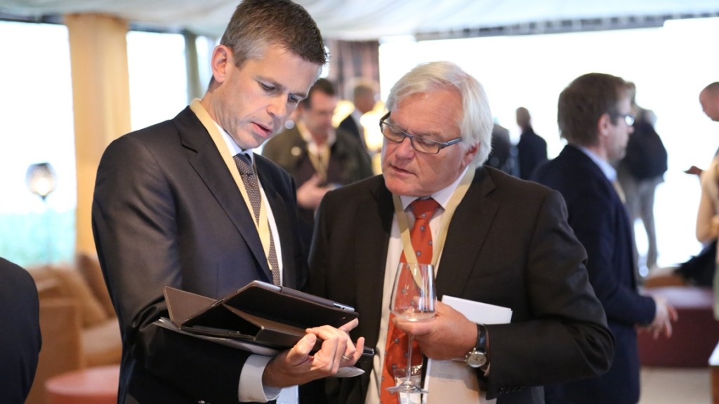
Dr. Erik Ranschaert (left – pictured here with Dr. Jan Schillebeeckx) from ‘s-Hertogenbosch, the Netherlands, will speak on mobile telera-diology with tablet computers in this Special Focus Session

Dr. Erik Ranschaert (left – pictured here with Dr. Jan Schillebeeckx) from ‘s-Hertogenbosch, the Netherlands, will speak on mobile telera-diology with tablet computers in this Special Focus Session

Read more…

ESR set to tackle personalised medicine and face economic realities in 2013

ECR2013_ECRToday_Monday_realities2013

In an interview with ECR Today, incoming ESR President, Professor Guy Frija explained how the ESR should address the challenges raised by the explosion of ‘omics’ data and the advent of personalised medicine, as well as how it should assess the impact of the financial crisis on the specialty.

ECR Today: What are your plans and ambitions regarding your presidency?
Guy Frija: The development of the ESR since its establishment in 2005 has been amazing: the ESR is now the biggest radiological society in the world, and its annual meeting, the European Congress of Radiology (ECR), is one of the best radiology congresses worldwide. All of this is the result of intense activity in several fields, such as education, research, professional challenges and publications, for the development of which the role of my predecessors was instrumental, along with the dedication of many of our colleagues. Therefore my first goal is to keep up this momentum and run the society appropriately, in accordance with its new statutes. However, life is challenging, and we will have to face many new issues in the coming year.

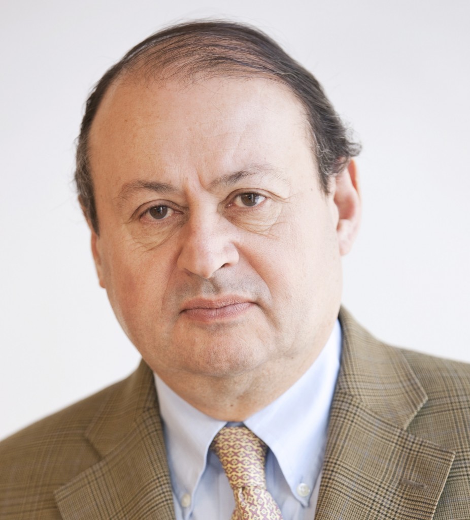
Professor Guy Frija is head of the imaging department at the Georges Pompidou European Hospital (Hôpital Européen Georges Pompidou, H.E.G.P.) in Paris.

Professor Guy Frija is head of the imaging department at the Georges Pompidou European Hospital (Hôpital Européen Georges Pompidou, H.E.G.P.) in Paris.

The explosion of ‘omics’ (such as metabolomics, proteomics, genomics) data is changing the face of medicine so considerably and rapidly that, if we don’t pay enough attention, our specialty could be threatened. Personalised medicine is not a just a dream, but an actual reality, and we have to adapt our specialty to this new paradigm. This year, DNA sequencing could become available for less than €750 (US$1,000), and in the UK, the Prime Minister David Cameron has launched a programme for the development of personalised medicine. The development of personalised imaging, which has already begun in oncology, should be taken further, and we have to appreciate its impact on education. I will ask two or three subspecialties to reconsider their training charters in light of these new developments. The data explosion related to these changes raises not only the problem of their management, but also of their interpretation in an integrated way: the imaging parameters should be included in this new data knowledge organisation, where knowledge modelling will become a major challenge. The establishment of imaging biobanks should be promoted along with adapted data privacy regulation.

Read more…

11
Mar 2013
POSTED BY
POSTED IN , ,
DISCUSSION 0 Comments

Imaging plays major role in fight against head and neck cancers

ECR2013_ECRToday_Monday_headneckcancer

Watch this session on ECR Live: Monday, March 11, 08:30–10:00, Room N/O
Tweet #ECR2013NO #SF16B

Organ-sparing surgery and radiation treatment such as intensity-modulated radiotherapy (IMRT) – often combined with chemotherapy – have increased the need for advanced imaging in the head and neck during pre-treament and post-treatment stages. Precision is vital as any tumour that remains undetected outside the treatment field could adversely affect the patients’ prognosis and survival, according to Professor Vincent Vandecaveye, from the department of radiology at the University Hospitals Leuven in Belgium.

It is important to spot any tumour recurrence as early as possible, especially in the post-treatment phase, in order give the patient the best possible chance of salvage treatment. The most common imaging methods in the head and neck area remain CT, MRI and PET-CT; each comes with its own advantages and disadvantages.

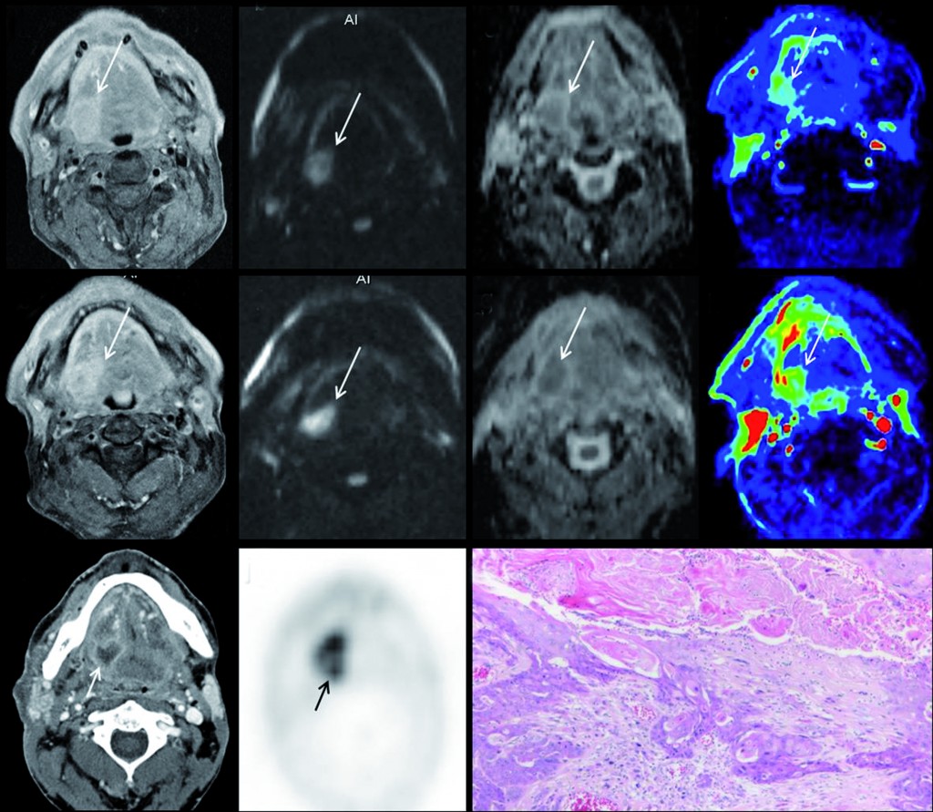
Multiparametric MRI for early treatment prediction of chemoradiation in oropharyngeal cancer:  Upper row is pre-treatment MRI of right base of tongue cancer (a=contrast enhanced T1 as anatomical correlate; b=native b1000 diffusion-weighted image; c= ADC-map; d=perfusion-map of IUAC). Middle row is 2 weeks during chemoradiation: same imaging sets, tumour volume will not help. No significant change in b1000, ADC nor perfusion-MRI indicate non-response and thus high risk of tumour relapse after end of treatment.  Tumour relapse at PET-CT 8 months after end of treatment, proven by histology (k). (Provided by Professor Vincent Vandecaveye)

Multiparametric MRI for early treatment prediction of chemoradiation in oropharyngeal cancer: Upper row is pre-treatment MRI of right base of tongue cancer (a=contrast enhanced T1 as anatomical correlate; b=native b1000 diffusion-weighted image; c= ADC-map; d=perfusion-map of IUAC). Middle row is 2 weeks during chemoradiation: same imaging sets, tumour volume will not help. No significant change in b1000, ADC nor perfusion-MRI indicate non-response and thus high risk of tumour relapse after end of treatment. Tumour relapse at PET-CT 8 months after end of treatment, proven by histology (k). (Provided by Professor Vincent Vandecaveye)

Read more…

10
Mar 2013
POSTED BY
POSTED IN ,
DISCUSSION 2 Comments

Advances in forensic imaging bring new opportunities for radiology

ECR2013_ECRToday_Sunday_Cover2

Watch this session on ECR Live: Sunday, March 10, 14:00–15:30, Room Z

The ability to spot pathological imaging findings among normal post-mortem signs of degeneration may not be a universal skill among radiologists, but it could prove to be a useful, if not essential one. As the reliability of modern forensic imaging rapidly improves, forensic pathologists are increasingly seeking the help of radiologists to examine bodies non-invasively, so they should be prepared to answer that call, according to experts who will speak in a Special Focus Session on ‘Advances in forensic imaging’ today. The development of techniques such as spiral volumetric CT and, more recently, MRI, have dramatically improved the ability of radiologists to determine causes of death and detect other crucial post-mortem signs, providing an invaluable service that can supplement, and in some cases replace, traditional autopsy.

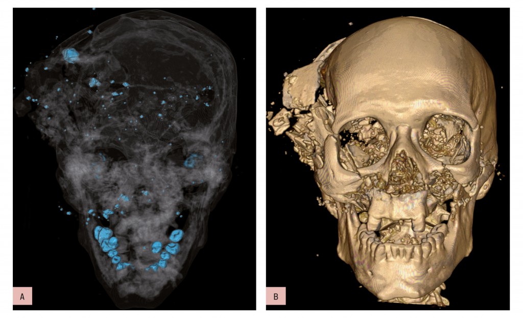
Bone and metal maximum intensity projection (MIP) reconstruction (a) and volume-rendered (VR) 3D-CT reconstruction (b). Homicidal death: characteristic bony and metallic fragments on the exit side of the skull, where the bullet caused a large loss of brain, leading to shattering of the skull.

Bone and metal maximum intensity projection (MIP) reconstruction (a) and volume-rendered (VR) 3D-CT reconstruction (b). Homicidal death: characteristic bony and metallic fragments on the exit side of the skull, where the bullet caused a large loss of brain, leading to shattering of the skull.

Read more…

10
Mar 2013
POSTED BY
POSTED IN ,
DISCUSSION 2 Comments