European Radiology: 25 years of excellence

Watch the European Radiology 25th Anniversary session live on ECR Online, Friday, March 3, 16:00–17:30, Room Z
#ECR2017 

Albert L. Baert, Adrian K. Dixon, Maximilian F. Reiser.

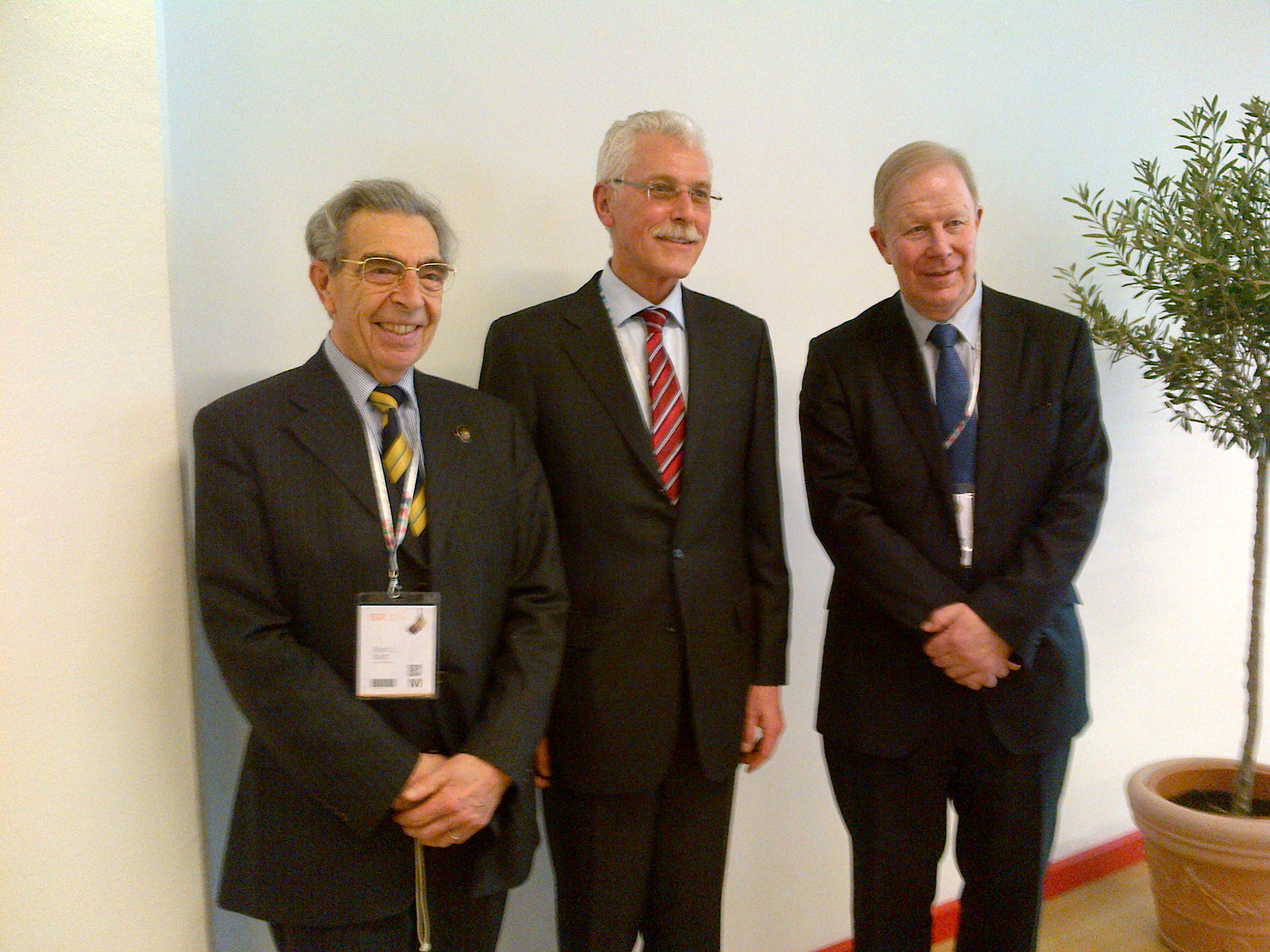
25 years of success for the three editors-in-chief of European Radiology: Albert L. Baert, Adrian K. Dixon, Maximilian F. Reiser.

The ESR’s flagship journal, European Radiology, celebrated 25 years of publication in 2016. We spoke to Prof. Albert L. Baert, the journal’s Editor-in-Chief from 1995 to 2007, Prof. Adrian K. Dixon, who headed European Radiology from 2008 until 2013, and our current Editor-in-Chief, Prof. Maximilian F. Reiser. Together, they gave us a comprehensive account of the journal’s development during the last quarter of a century.

 ECR Today: How have you seen the journal develop during the past 25 years?

Albert L. Baert: During this time period the number of published articles has increased spectacularly from 60 articles per year in the first issues to more than 400 per year now. For example, no less than 4,674 pages were published in 2016! European Radiology is now one of the most widely distributed and read scientific journals in the world. Simultaneously, the scientific level of the contents has improved steadily over the years as proven by the current excellent Impact Factor ranking.

Read more…

02
Mar 2017
POSTED BY
POSTED IN ,
DISCUSSION 0 Comments

Facial genetics and forensics take centre stage at ECR

Watch this session on ECR Live: Friday, March 3, 10:30–12:00, Room B
#ECR2017

Forensics and facial genetics will be in the spotlight at ECR 2017 as the ‘ESR meets Belgium’ session offer a look at medical imaging’s most original contributions to healthcare and crime investigation.

Advanced decomposition of brain MRI or facial 3D surface images into modules, tailored for associations with underlying genetic variations.

Dr. Peter Claes from Leuven, Belgium, will present his work in facial genetics in a session titled ‘Imaging genetics and beyond: facial reconstruction and identification’.

Claes is a senior research expert in the Medical Image Computing research group of the Processing of Speech and Images division of the Electrical Engineering department at Leuven Catholic University.

He uses CT, MRI and 3D surface imaging modalities to grasp the link between people’s appearance and underlying genetic variations.

“Your appearance is genetically driven. In families there’s a strong link, even more so between identical twins, who share the same DNA profile and almost the same face. Physical features also influence your brain, the way you think. A lot of facial characteristics are shared, for example in Down’s syndrome patients, who present with the same features whether they are European or Asian,” he explained.

The link between genetic disorders and facial genes has been of interest to scientists for a while, but research is slow and tedious.

Claes became interested in facial genetics after working in craniofacial morphometrics to help correct morphological abnormalities and anomalies, and in craniofacial reconstruction to identify victims.

To help decipher facial genetics, Claes uses the computer-based craniofacial reconstruction programme he developed for victim identification, and combines 3D surface processing, statistical modelling, analysis, mapping and prediction techniques. He has also created an array of algorithms and software for investigators who plan to use 3D facial datasets. Last year, he also co-organised the first international workshop on facial genetics in London.

Read more…

Celebrating Ten Years of ESOR

Guest article by ESOR Director, Prof. Nicholas Gourtsoyiannis

Nicholas Gourtsoyiannis

Nicholas Gourtsoyiannis is the Educational & Scientific Director of the European School of Radiology (ESOR) and chairman of the ESR’s ESOR Committee.

The European School of Radiology (ESOR) has completed ten years in action. Ten challenging and rewarding years of unfailing commitment and continuous investment in radiological education in Europe. Ten years of envisioning, engaging, delivering, teaching, tutoring, nurturing, and adding value to radiology.

The three main goals of ESOR are still to assist in harmonising radiological education throughout Europe, by supporting the implementation of the European Training Curricula (ETC); to build a genuine and firm interest in subspecialisation in radiology; and to raise the scientific profile of radiological education in Europe and worldwide.

The past ten years of ESOR have been marked by an outstanding growth in a wide range of modular activities, including foundation and advanced courses, teach-the-teachers and visiting professorship programmes, visiting schools, seminars, tutorials, preparatory courses for the European Diploma in Radiology (EDiR), scholarships, exchange programmes for fellowships and full one-year fellowships. So far, ESOR has delivered structured continuing education to almost 17,000 residents and board-certified radiologists worldwide, through 800 programmes.

Read more…

02
Mar 2017
POSTED BY
POSTED IN ,
DISCUSSION 0 Comments

Education central to improving imaging data quality in oncology clinical trials

Watch this session on ECR Live: Thursday, March 2, 16:00–17:30, Room X
#ECR2017

Imaging data is key in multicentre clinical trials for cancer research but quality control is currently a major impediment, bringing the validity of the trials into question and potentially impacting on the quality of drugs put on the market, a panel of experts will argue today in a session held by the ESR and the European Organisation for Research and Treatment of Cancer (EORTC) at the ECR.

Imaging is increasingly contributing to cancer research thanks to the development of innovative techniques that depict functional and molecular processes. In most oncological clinical trials, imaging is now the primary criteria used to evaluate progression of disease or efficiency of the drug being tested.

The best way to obtain valuable imaging measurements is to involve the imagers who take part in these trials and educate the clinician investigators, experts will explain in the session.

When it comes to imaging in cancer research, a number of issues take centre stage. Difficulties associated with integrating imaging biomarkers into trials have been neglected compared with those relating to the inclusion of tissue and blood biomarkers, largely because of the complexity of imaging technologies, safety issues related to new contrast media, standardisation of image acquisition across multivendor platforms and various post-processing options available with advanced software, as reported recently in The Lancet by the EORTC and leading researchers.

Read more…

01
Mar 2017
POSTED BY
POSTED IN ,
DISCUSSION 0 Comments

Eminent neuroradiologist to give honorary lecture at ECR 2017

Watch this session on ECR Live: Thursday, March 2, 12:15–12:45, Room A
#ECR2017

In recognition of his significant impact on the field of neuroimaging and his service to organised radiology, Professor Mauricio Castillo from Chapel Hill, NC, United States, has been invited to deliver the Wilhelm Conrad Röntgen Honorary Lecture ‘Dissatisfaction, burnout and inequality: three major challenges in radiology’ at ECR 2017.

Professor Mauricio Castillo from Chapel Hill, United States

Professor Mauricio Castillo from Chapel Hill, United States, will deliver the Wilhelm Conrad Röntgen Honorary Lecture today at 12:15 in Room A.

Mauricio Castillo is the James H. Scatliff distinguished professor of radiology, chief and programme director of neuroradiology at the University of North Carolina in Chapel Hill. He is currently president of the American Roentgen Ray Society.

Originally from Guatemala, Prof. Castillo completed his radiology and neuroradiology training at the University of Miami School of Medicine, Jackson Memorial Medical Center, and Emory University School of Medicine, Affiliated Hospitals in Atlanta respectively.

His specialty and subspecialty choice was in large part motivated by his mentors.

“When I was a medical student, I was fortunate to work in the only teaching hospital that had a CT scanner and two board-certified radiologists. I was very impressed by the impact of imaging in patient care and by the way these two professionals used their large base of knowledge of medicine to generate a differential diagnosis based on imaging findings. Later on, while I was a resident, I developed a close relationship with Dr. Robert Quencer, which led me to become a neuroradiologist. One should never underestimate the influence we may have on our trainees and the influence our mentors have had on us,” he said.

Prof. Castillo’s research interests include paediatric neuroimaging, application of new imaging techniques and medical literature editing. Read more…

Image-guided interventions: a key pillar in cancer care

Watch this session on ECR Live: Wednesday, March 1, 16:00–17:30, Room F2
#ECR2017

 There is hardly any area of hospital medicine where interventional radiology (IR) has not had some impact on patient management. The range of conditions that can be treated using interventional radiology techniques is continually expanding.

In today’s session, experts will provide an insight into image-guided interventions in oncology with a particular focus on illustrating the importance of quality assurance in image-guided oncological interventions and their effect on treatment outcomes.

In recent years, IR has played a vital role in the field of oncology, and alongside medical, surgical and radiation oncology it constitutes a key pillar in cancer care. Vascular and non-vascular procedures such as transarterial chemoembolisation, radiofrequency ablation (RFA), microwave ablation, radioembolisation, cryoablation and high-intensity focused ultrasound (HIFU) are delivered locally, minimise damage to nearby tissue and avoid the systemic side effects of chemotherapy.

Colorectal lung metastasis before CT – guided microwave ablation.

For the interventional radiologist providing oncologic therapies it is essential to understand the rapidly changing field of oncology and to have a broad knowledge of oncologic diseases and available therapies to treat them. Radiologists providing image-guided interventions in oncology have an outstanding understanding of imaging as well as a diversity of interventional skills. However, they lack formal training in oncology and an understanding of chemotherapy and radiotherapy, according to Prof. Andy Adam from the Department of Radiology at Guy’s and St. Thomas’ Hospital in London. Read more…

Prominent Brazilian radiologist to shed light on Zika virus imaging at ECR 2017

In recognition of her major achievements in neuroimaging and advancement of the field, Professor Maria de Fátima Viana Vasco Aragão from Recife, Pernambuco, Brazil, will present the Guest Lecture ‘Breaking News from Latin America: How to recognise Zika virus infections on imaging studies’ at ECR 2017.

Maria de Fátima Viana Vasco Aragão is professor of radiology at the Maurício de Nassau University and Scientific Director of the Multimagem Diagnostic Centre in Recife, Brazil. She is also financial director of the Diagnostika Endoscopy and Radiology Clinic in Recife.

Prof. Vasco Aragão received her medical degree from Pernambuco Federal University in 1987 and completed her residency in radiology at Ribeirão Preto Faculty of Medicine, University of São Paulo. She did fellowships in CT and MRI and in neuroradiology at the Med Imagem Beneficencia Portuguesa, São Paulo, and later completed a research fellowship at Mount Sinai Hospital Center in New York, US.

She always knew she would become a doctor and chose radiology because she recognised early on the field’s value in diagnosis and treatment.

Maria de Fátima Viana Vasco Aragão is professor of radiology at the Maurício de Nassau University and Scientific Director of the Multimagem Diagnostic Centre in Recife, Brazil.

Maria de Fátima Viana Vasco Aragão is professor of radiology at the Maurício de Nassau University and Scientific Director of the Multimagem Diagnostic Centre in Recife, Brazil.

“My mother is a retired biologist and biology teacher. I have never forgotten the moment she taught me what a cell was. In my family, educators predominated and my great aunt was the first woman in Pernambuco, my home state, to graduate from medical school, in 1935. Perhaps on account of this family background I made up my mind to become a doctor. As an intern student of medicine, I was very impressed when I took my patients to be examined by CT and US. Even though there was only one CT scanner in my city, I succeeded in having all my intern patients examined, when indicated. My tutors were impressed by the swiftness with which cases were resolved. Thus I learned that radiology not only helps to quickly define diagnosis in most patients, but also provides a clear prognosis for the most adequate treatment. At the end of the course I began considering the possibility of specialising in radiology, even though 29 years ago, for most people, a radiologist was not a doctor. Shortly after graduation, I sat for a test and won a place in radiology at the first attempt, and radiology has been my passion ever since. I would like to say that I am grateful for having had great mentors, including the neuroradiologist Dr. Sérgio Santos Lima, who was my director during my fellowship in the Med Imagem Beneficencia Portuguesa, and the head and neck radiologist, Dr. Peter Som, who was one of my directors during my research fellowship at Mount Sinai Hospital Center New York,” she said.

Read more…

ESR and Materialise unveil Medical 3D Printing Experience Center together at annual meeting

3D technology has been transforming the face of healthcare for over 20 years. 3D-printed anatomical models are increasingly being used for evaluating treatment approaches, planning complex procedures and improving the training of medical professionals. And that’s just the start.

The ECR presents a great opportunity to explore the contributions and benefits that Medical 3D Printing brings to healthcare at the 3D Printing Experience Center in the Entrance Hall. To bring this experience to life, the ESR is collaborating with Materialise, as the backbone of the 3D printing industry.

3D-printed skull

You will experience the workflow interactively – from medical images to accurate 3D anatomical model, explore all the different clinical applications of Medical 3D Printing and learn the first steps of setting up a 3D lab in your hospital.

But why exactly is Medical 3D Printing generating so much interest amongst radiologists?

Read more…

23
Feb 2017
POSTED BY
POSTED IN ,
DISCUSSION 0 Comments

The best unused cases submitted for the popular “Know Your Calcifications” interlude at ECR 2016

ECR2016_banner_blog-focus

Dear Friends,

Over the last couple of years, one of the last sessions at the ECR has always covered 20 interesting cases from various subspecialties, which the audience are asked to solve in an interactive way to broaden and update their knowledge.

In between, the very best submissions from the global radiological community have been presented in an interlude lecture. The best submission has always been awarded with a prize and a certificate.

Due to time limits, only a small number of submitted cases can actually be shown onsite, but the session’s rising popularity has resulted in increasing numbers of submissions of excellent quality. This is why we would like to give our submitters the opportunity to reach a broader audience by posting the best cases here on the ESR Blog.

Read more…

ECR 2016 Cases of the Day Winners

ECR2016_banner_blog-update

The winners of the ECR 2016 Cases of the Day Quiz are as follows:

S.A. Sohaib; Sutton/UK
Kemal Kara; Istanbul/TR
Ersin Ozturk; Istanbul/TR
Bertram Feil; Zurich/CH
Miguel Nogueira; Marinha/PT
Yu Kuo; Taipei/TW
Filip M.H.M. Vanhoenacker; Antwerp/BE
Bilal Battal; Ankara/TR
Floor van der Wolf – de Lijster; Sneek/NL

To view the cases please click here.

Congratulations to all winners!