Following the usual pattern of an oral examination (mixed cases from all the subspecialities), I challenge you with the following neuro case, below.
Look out for the answer on Thursday. Good luck!
This week’s patient is a 61-year-old immunocompetent male with progressive disorientation and general weakness
1. Metastases
2. Pyogenic abscess
3. Tuberculosis
4. Multiple Sclerosis
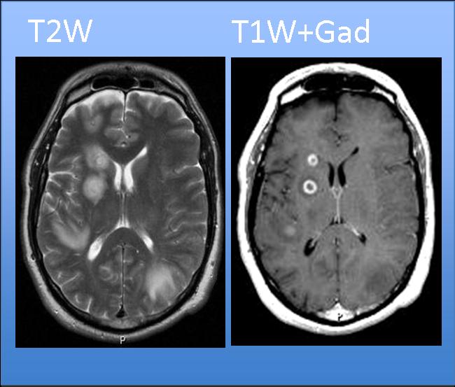
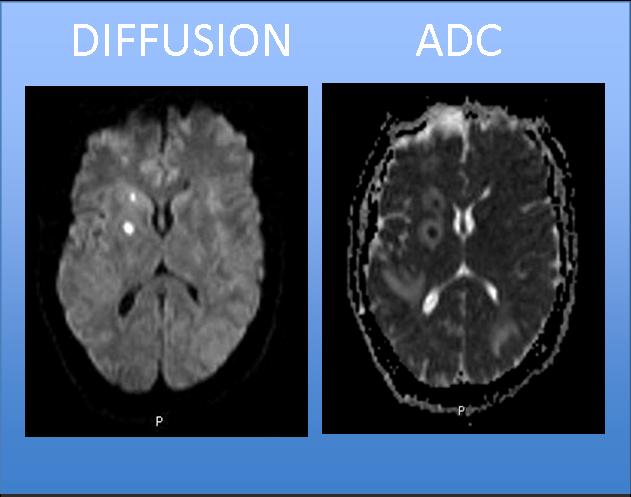
Findings: multiple intraparenchymal hemispheric lesions, with a peripheral rim, oedema and ring enhancement (yellow arrows). Low ADC values on diffusion images (red arrows).
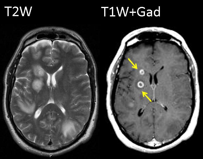
Fig. 4
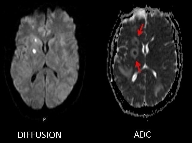
Fig. 5
Although the most common ring enhancing lesions in immunocompetent adult patients are metastases, the low ADC values are very suggestive of brain abscesses. Occasionally, metastasis and MS plaques may demonstrate low ADC values inside the ring enhancing areas; but it is an uncommon finding
Final diagnosis: Pyogenic brain abscesses
Brain abscesses typically demonstrate a complete ring of enhancement with a smooth inner and outer margin and reduced diffusion coefficient, and therefore low signal intensity on the apparent diffusion coefficient map. On MR imaging, tuberculous abscess can present the same findings as pyogenic abcesses. Clinical history, clinical presentation and laboratory tests are the most important clues to make the diagnosis.
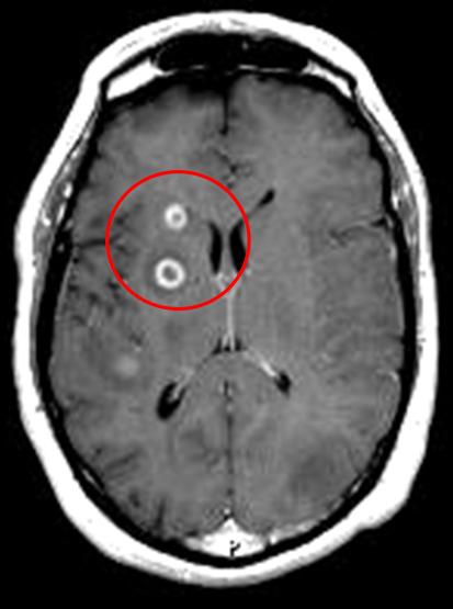
Fig. 6 Continuous ring of enhancement
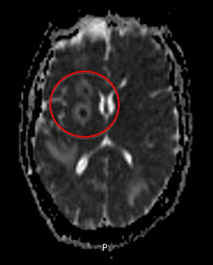
Fig. 7 Low ADC
Differential diagnosis should include metastases, specially in adults and immunocompetent patients. It is important to know the prior clinical history of the patient to make the diagnosis. Ring enhancement and restricted water diffusion also might be found in brain metastases.
Fig. 8
Patients with MS may present with multiple ring enhancing lesions. The most characteristic finding is an ‘incomplete ring’ of enhancement (arrow). MS plaques show a discontinuous ring that is not demonstrated in cases of metastasis or abscesses.

Fig. 9

Follow Dr. Pepe’s advice
- Differential diagnosis of brain ring enhancing lesions should include metastases, abscesses, tuberculosis, MS.
- Low ADC values are highly predictive of abscess, although they are not 100% specific.
- An ‘incomplete ring’ of enhancement suggest demyelination.
Suggested reading. Patterns of Contrast Enhancement in the Brain and Meninges.James G. Smirniotopoulos. RadioGraphics 2007; 27:525–551
Case prepared by Laura Oleaga MD


At least three lesions that restrict in diffusion and have ring heighten after contrast administration. Vasogenic edema is also present.
Option 2 : Pyogenic abscess.
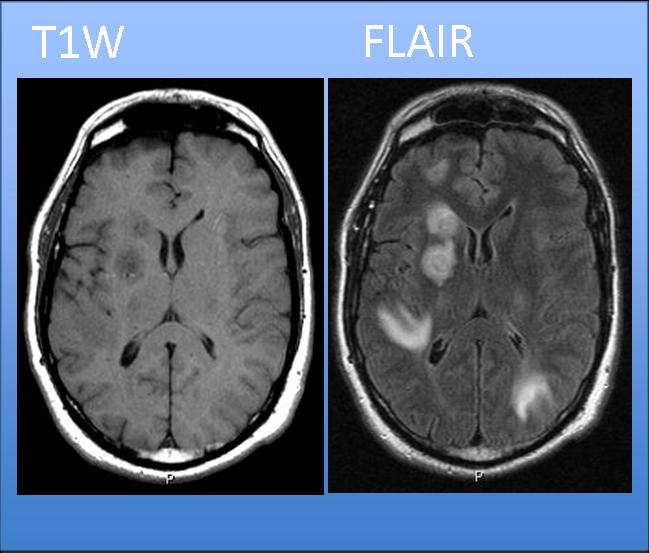
In T2-FLAIR hyperintense areas are seen extended in the subcortical white matter of right temporal and left occipital lobes, maybe in relation with encephalitis.
Pyogenic abscess
I agree. Option 2: pyogenic abscess.
TB for immuno competent
Abcese pyogenic.
Pyogenic abscess.
Does he have fever? It seems to be pyogenic abscess.
2. Pyogenic abscess
PYOGENIC ABSCESS , ENCAPHALITIS
Tuberculosis
Tuberculosis (with vasculitis ??)
Pre and post contrast MR axial images showing multiple ring enhancing lesions in right basal ganglia with surrounding edema. These lesions show central hypointensity on T1 and hyperintensity on T2W with peripheral rim appearing isointense on T1 and hypointense on T2W images. The central core shows diffusion restriction.Non enhancing lesion with similar character on T2W is also seen in right frontal region.
Areas of white matter edema are seen in right posterior temporal and left occipital regions with nodular enhancing lesion in right posterior temporal region. no appreciable enhancing lesions are seen within these areas. no abnormal meningeal or ependymal enhancement is seen. no hydrocephalous seen. the differentials can be -pyogenic or tubercular abscesses.In view of deep seated lesions, lesions in different stages and hypointense rim on T2W images-my first possibility is tubercular.
I agree completely with vishal…however, common things being common, I would place pyogenic abscesses before TB.
I agree with both of you. Lacking clinical information, go for the most common diagnosis
Thanks for the feedback and effort you put in this!
Ascessi da piogeni: dd con ascessi TBC.La presenza di forme “immature” di cerebrite( aree iperintense a limiti sfocati) depone per ascessi batterici.Gli eventuali test , negativi, per TBC( Mantoux eB-Quantiferon nel siero) ne avvalorano la diagnosi.
You all did very well! Keep up and you will pass the Diploma easily.