
Dear Friends,
This year I plan to show only chest cases, emphasising the diagnostic approach to basic patterns in the plain film. Hopefully, this monographic approach will help you with the diploma examination.
Cases will be posted every other Monday and answers will be given on Friday.
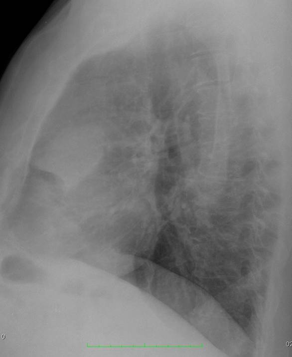
Radiographs (below) of the first case belong to a 57-year-old man, asymptomatic. Study them carefully, leave your opinions in the comments section, and look out for the answer on Friday.
Diagnosis:
1. Thymoma
2. Teratoma
3. Mediastinal fat
4. Can’t tell
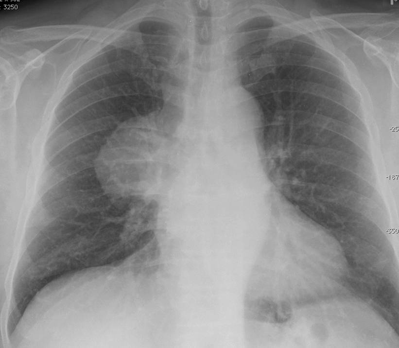
57-year-old man, asymptomatic
57-year-old man, asymptomatic
Click here for the answer
Findings: chest radiographs show an anterior mediastinal mass (arrows). The obvious choice is either thymoma or teratoma. However, it is important to remember that in chest plain films, all mediastinal masses have similar densities and fat cannot be distinguished from soft tissue. Therefore, the correct answer should be no. 4:
Can’t tell
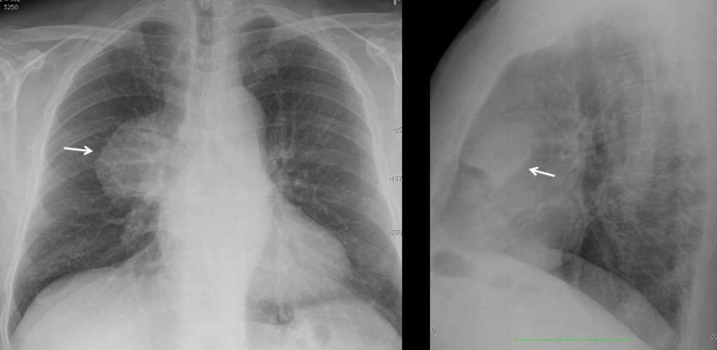
Fig. 1
Axial and coronal CT images show a well-defined fatty mass (A,B arrows), with no internal vessels.
Final diagnosis: mediastinal fat/mediastinal lipoma
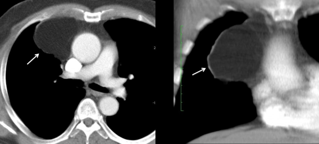
Fig. 2
This case is shown to illustrate that all mediastinal masses have similar density in plain films. To determine their radiographic density, CT must be performed.
The role of plain films in mediastinal masses is:
1. To discover them
2. To localise them in a mediastinal compartment, to narrow down the differential diagnosis
Once these steps are taken, it is imperative to do cross-sectional imaging (usually CT) to confirm the location of the mass, its radiographic density, and response to contrast enhancement.

Follow Dr. Pepe’s advice:
- The role of chest radiography in mediastinal masses is to identify and locate them.
- Mediastinal masses are characterised using enhanced cross-sectional imaging.
- The great imitators are vascular structures, mediastinal fat, and oesophageal disease.


Opacità nel mediastino anterosuperiore: regola delle 4 T( Tiroide-Timo-Teratoma-T linfoma).Tiroide esclusa perché manca il raccordo con un prolungamento intratoracico di una tiroide).Timoma ,T.da residui embrionari e Linfoma per mancanza di clinica e-o laboratorio).Rimane grasso mediastinico soprattutto se ha fatto terapia steroidea.Farei solo una ecografia perché la sede lo consente(evitando la Tac).
I think that the lesion is situated in the anterior mediastinum (not anterosuperior) and that it can be either a thymoma, a teratoma or a thymoma but not a thyroid lesion, because the latter would be situated in the middle mediastinum.
I choose 1. Thymoma
because it is the most common tumor in the anterior mediastinum, it is often asymptomatic and the mean age of presentation is 50 years
See my answer to Gus, below
(By the way, I wrote taken instead of taking. Apologies)
the mass is in the anterior mediastinum and i think is more dence for mediastinal fat.could be a cystic mass or a mixt density mass. there is not calcified and there is not mass efect on the adjacent structures.
thymoma is the best choise
but the most sophisticated answer is i can’t tell.
CT can
OK, but will you do in real life? Or when taken the exam?
there is fat on the right cardiophrenic angle. maybe on right superior mediastinum too?.
dr pepe after exam we have party:)
Sure, I will bring the booze
I would say thymoma. differential would be teratoma, but the age of the patient doesn’t really fit.
I think it’s in the anterior mediastinum, well circumscribed, large. I think a Tymoma, or maybe a pericardial cyst in a less likely location.
…anche una cisti dermoide, potrebbe essere la risposta giusta.
Before posting the answer tomorrow, I would like to ask you if it is possible to make an specific diagnosis of a mediastinal mass in the plain film.
….per me non è possibile….si può dire che la massa è compatibile in prima istanza diagnostica con….e poi confermarla con una TAC( se si ha una sonda ecografica , si può completare con questa)
Correct! Therefore, the best answer is 4. Cannot tell
The message is that any mediastinal mass must be evaluated with cross-sectional imaging (usually, but not only, CT)
…corretta la mia diagnosi: Bari-Barcellona 2 a 0. Reti:Casebook Pepe’s 41 e Prof.Caceres casebook 71!!!!
Nella mia carriera mi era già capitato di vedere un’immagine simile e il medico radiologo lo aveva refertato per massa di grasso a livello mediastinico..consigliando però alcune scansioni TC mirate..
An ant. mediastinal mass, of well-defined outline, I think
the best answer is 4 , because further evaluation is needed by CT scan.
[…] Most mediastinal lesions are discovered in the PA radiograph. Broadly speaking, they look like soft-tissue ‘bumps’ in the mediastinum (Fig. 1), identified by the typical pregnancy sign (Diploma Casebook case 95). Since the great majority have no specific characteristics, the role of the chest radiograph is to place the lesion in one of the mediastinal compartments (see Diploma Casebook case 41). […]