ESR Rising Stars Speakers at ECR 2012
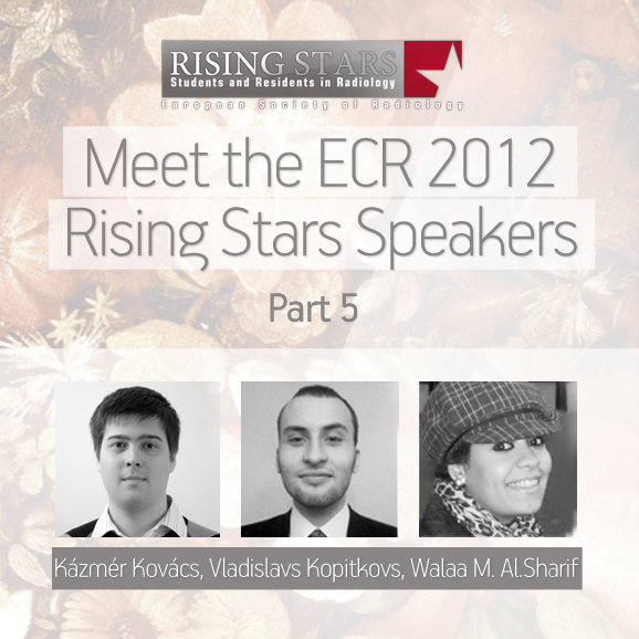
Welcome to the fifth installment of our series introducing our Rising Stars, who submitted the best student abstracts and have been invited to present their work at ECR 2012 in front of a real congress audience. Each speaker will be announced individually on the Rising Stars Facebook page, with a more detailed look, including their full abstracts, here on the ESR Blog. Our next three speakers are Kázmér Kovács, Vladislavs Kopitkovs and Walaa M. Al.Sharif:
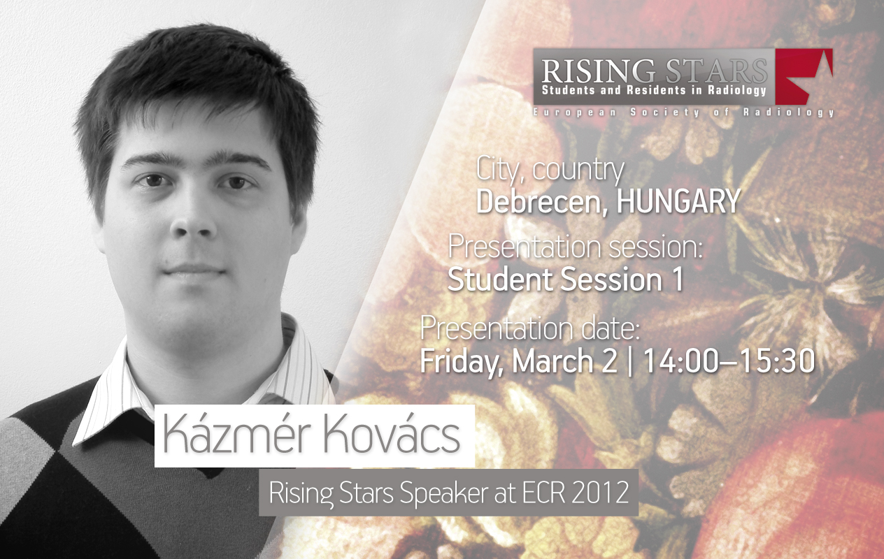
ECR 2012:
Student Session 1 | March 2, 14:00 – 15:30
CV (excerpt):
Education/Profession:
Since 2006 Studying Medicine at the Faculty of General Medicine, Medical and Health Science Center, University of Debrecen, Hungary
Extracurricular activities:
Since 2010 Editor-in-chief for the university magazine Medikus Lap
Summer 2011: Participant in a medical imaging research group in the Department of Biomedical Laboratory and Imaging Science, University of Debrecen.
Abstract:
Introduction: Our aim is to design a system that will help master the daily diagnostic radiological routine. This, of course, will not replace consultation with the specialist, but should make it easier to recognise diseases. Discussion: The programme is basically a computer simulator for localising and identifying pathologies. After the evaluation of MRI, CT, US, or X-ray images, the device checks if the user’s answer is correct. This would be based on predefined diagnoses by imaging specialists. Initially, it would be necessary to construct a large dataset of correct diagnoses and detailed, good quality patient records (request forms and images). Plain radiographs and particularly cross sectional CT and MRI images can be easily applied to this learning method, already there are tools available for this. We suggest an alternative, easy-to-use way to perform US scans, the acquisition and reconstruction of ultrasound. In order to allow for the computer-assisted simulation of a US scanning procedure, US scans should be performed with 3D-guidance: storing spatial information regarding the transducer’s position and orientation. Using a 3D controller device in this virtual space, the user would be able to perform simulated US acquisitions. The database would be a result of international collaboration, but local radiology units could also create their own material based on their own patient database. The simulator suggests different training modes based on the imaging techniques and the disease’s distribution. To encourage learning and review, scientific papers, or unified radiological eLearning material (such as eurorad.org) could be attached to cases. Conclusion: We assume that this simulator could revolutionise the training of radiological image recognition. It provides a supervised knowledge basis, especially for daily diagnostic routine.
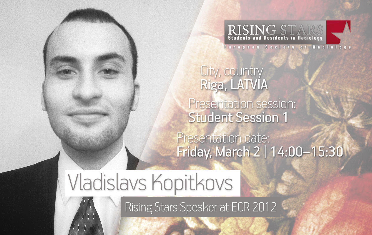
ECR 2012:
Student Session 1 | March 2, 14:00 – 15:30
CV (excerpt):
Education/Profession:
Since 2008 Student at Riga Stradiņa University, Latvia
Extracurricular activities:
Since 2011 Vice-President of the Latvian Medical Students Association
Since 2011 Volunteer at the Radiology Department, Riga Eastern University Clinic
Abstract:
Just like today, in the year 2051 radiology will remain one of the most rapidly developing medical specialties, which will continue to offer new research methods to other medical fields, giving them the capability to increase the accuracy of their diagnoses. The widespread usage of imaging methods will noticeably decrease their cost and provide job vacancies for radiologists. Radiological procedures will become significantly safer for both the patients and medical personnel. Hypoallergenic and non-nephrotoxic contrast materials will be produced. I can clearly imagine the future, where radiological equipment will become mobile, which will finally give us the option of taking crucial diagnostic examinations to the patients who are not able to reach medical facilities themselves. In acute cases, radiological imaging will be available at the site of various emergencies, battlefields and natural disasters. Routine full body scanning will even allow for prompt automatic diagnosis with any organ size and morphological changes or neoplasms in comparison to previous scans. A unified database of body scans will allow any physician in the world to access medical records. Automatic mass screening procedures will allow for the detection of diseases among populations and newly arrived tourists, giving us a chance to take preventive measures against the spread of disease. The person at risk will be detected and given medical treatment. There will be a compulsory radiology course for all other specialised physicians, in terms of their specific field. Doctors will be encouraged to carry out simple non-invasive diagnostic procedures on the spot and request a radiologist’s assistance only if complex procedures are required. That is my view of the future of radiology.
ECR 2012:
Student Session 4 | March 3, 10:30 – 12:00
CV (excerpt):
Education/Profession:
Bachelor of Diagnostic Radiology, King Abdul-Aziz University, Jeddah, Saudi-Arabia
MSc programme in Magnetic Resonance Imaging, University College Dublin, Ireland
Extracurricular activities:
2009: 6-month rotation in radiology department, King Fahad Arms Forces Hospital, Jeddah, Saudi-Arabia
2009-2010: Internship in MRI at King Abdul-Aziz University hospital, Jeddah, Saudi-Arabia
Abstract:
MRI is a major tool in the field of diagnostic imaging. However, there are several factors which could affect MR image quality and they appear for a variety reasons. Potential sources of which include: hardware characteristics, intrinsic tissue properties, and sequence parameters. Therefore, it is essential to choose the pulse sequence with accurate imaging parameters, because those represent a vital factor in diagnostic techniques. Furthermore, optimum image is a compromise between the SNR, CNR, Spatial Resolution, and their own affected factors, and many artefacts could appear because of them. However, in some cases it could be corrected and mitigated to improve the image quality. Methodology: In this study we have quantitatively analysed the impact of alteration in the imaging parameters of image quality in T2W – FSE- axial plan MR imaging. In this case FSE was used as the baseline scan time for FSE was faster, because several sequences were acquired from the same volunteer during one session. 1) Acquire the baseline T2W FSE sequence – standard optimised parameters. 2) Run the sequence as per 1) above, except reduce the TR. 3) Run the sequence as per 1) above, except reduce the TE. 4) Run the sequence as per 1) above, except reduce the number of acquisitions. 5) Run the sequence as per 1) above, except reduce the slice thickness. 6) Run the sequence as per 1) above, except reduce the FOV. 7) Run the sequence as per 1) above, except reduce the matrix. Result: There are several factors which affect MR image quality. However, there are numerous correction methods which could improve the MR image resolution to facilitate evaluation and diagnosis of abnormalities. These methods include choosing sequences, scanning procedure, hardware, and parameters .


check this link, cheap coach handbags at my estore