Interview: Prof. Boris Brkljačić, professor of radiology and Vice-Dean at the University of Zagreb School of Medicine, Croatia

This year, the main theme of the International Day of Radiology is breast imaging. To get some insight into the field, we spoke to Prof. Boris Brkljačić, professor of radiology and Vice-Dean at the University of Zagreb School of Medicine, Croatia, and Chairman of the ESR Communications and External Affairs Committee.
European Society of Radiology: Breast imaging is widely known for its role in the detection of breast cancer. Could you please briefly outline the advantages and disadvantages of the various modalities used in this regard?
Boris Brkljačić: Mammography, ultrasound and MRI are three modalities used for the detection of breast cancer. Mammography has been used for many decades, and the introduction of full flat panel digital mammography has enabled image acquisition with a lower radiation dose, and other advantages in image processing and biopsies. Mammography is used widely in breast cancer screening and has been validated through decades of screening. It is also the initial imaging method in women older than 40 and it enables the detection of microcalcifications, the early signs of ductal cancer in situ, and the majority of breast cancers, depending on the radiographic density of the breast. It can also be used to guide biopsy of microcalcifications. The denser the breasts are, the lower the sensitivity of mammography in detecting breast lesions, which is the disadvantage of mammography. The new mammographic method, digital tomosynthesis, improves the detection rate of cancer in dense breasts. Mammography exposes patients to radiation and is therefore not recommended in young women because their breasts are very radiosensitive.

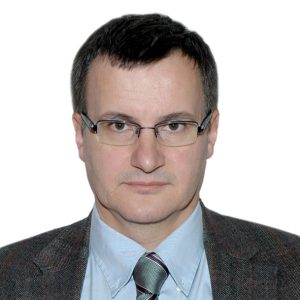
Prof. Boris Brkljačić, Professor of Radiology and Vice-Dean at the University of Zagreb School of Medicine, Croatia, and Chairman of the ESR Communications and External Affairs Committee.
Ultrasound is an imaging method that provides images based on the acoustic properties of tissues. The blood flow in lesions can be analysed by colour Doppler ultrasound, and elasticity of lesions can be analysed and quantified by sonoelastography. The advantage of ultrasound is that it is completely harmless; it does not expose patients to radiation, and is an excellent method for the guidance of biopsies of all sonographically visible lesions. Ultrasound can demonstrate cancers that are not visible in mammographically dense breasts, and is the complementary imaging modality to mammography, both in diagnosis and in screening. Some U.S. states legally oblige physicians to inform women about mammographic density and advise them of additional methods of examination in dense breasts. Among many advantages in ultrasound technology are the automated whole-breast ultrasound systems that have recently been introduced to the market. The disadvantage of ultrasound is that it increases the number of false-positive findings.
Magnetic resonance imaging (MRI) of the breast has gained considerable importance over the last two decades and is used more and more in breast imaging. It is used in high-risk screening, in the detection of occult cancer with positive lymph nodes, and in the evaluation of implants, and it is the best method for detecting the presence of and assessing the distribution and extent of cancer. It can also be used to monitor the success of neoadjuvant chemotherapy, and is an excellent method for looking for residual cancer or recurrence after treatment. MRI is relatively expensive and time consuming, although abbreviated MRI protocols have recently been introduced.
For treatment planning and monitoring it is very important to know the exact type and grade of cancer, and its immunohistochemical profile. Image guided biopsy is crucial in relation to that, and all imaging methods enable precise, image-guided biopsy to obtain an adequate sample from the breast cancer and other breast lesions.
ESR: Early detection of breast cancer is the most important issue for reducing mortality, which is one reason for large-scale screening programmes. What kind of programmes are in place in your country and where do you see the advantages and possible disadvantages?
BB: A mammographic breast cancer screening programme has been in place in Croatia since October 2006, and it is a national programme, covering the whole population of women in the age group 50–69. The advantages and disadvantages of this programme are similar to other screening programmes. The Croatian programme has some additional problems because, in order to provide coverage of the whole country, many screening units were set up, but the level of breast imaging experience among the staff at those units tends to vary. The distribution and renewal of equipment has also not always followed the most rational plan.
ESR: Do you know how many women take part in the Croatian screening programme (percentage)? Do patients have to pay for this?
BB: In the Croatian national mammographic screening programme the annual attendance rate so far has been 57–63%. The regional differences are quite pronounced; some counties have attendance rates in the range of 70–80% while in the others the rates are much lower. The patients do not have to pay for screening since the costs are covered by the healthcare budget, i.e. the national medical insurance fund pays for all the costs.
ESR: The most common method for breast examination is mammography. When detecting a possible malignancy, which steps are taken next? Are other modalities used for confirmation?
BB: The most common clinical scenario is that ultrasound is performed after mammography, and ultrasound-guided core biopsy would then be performed to diagnose cancer and assess the exact type of cancer. If the lesion is visible only on mammography, then stereotactic, mammographically-guided core biopsy would be performed. In many instances breast MRI is performed before the treatment to assess the size, distribution and extent of cancer.
ESR: Diagnosing disease might be the best-known use of imaging, but how can imaging be employed in other stages of breast disease management?
BB: The role of imaging after surgery is very important. Mammography, ultrasound and MRI are used regularly in the follow-up of the patient, and these modalities are crucial in the detection of residual cancer or recurrent cancer. The current trends are that less surgery is being performed, and smarter, more targeted therapies are being introduced. Chemotherapy before surgery is more common, and patients are being selected for neoadjuvant chemotherapy instead of primary surgery on the basis of imaging findings. Also, the success of such therapy is monitored using imaging, primarily MRI.
ESR: What should patients keep in mind before undergoing an imaging exam? Do patients undergoing radiological exams generally experience any discomfort?
BB: Patients undergoing mammography usually experience a certain degree of discomfort, because the breast is compressed during the examination. In my experience, most women do not find this very disturbing. Ultrasound examinations are usually quite easy for the patient, without discomfort. During MRI examinations, patients need to be still, lying in the prone position, with breasts positioned within the dedicated breast coils. This position is not very comfortable. Some patients are also unable to tolerate MRI examinations due to claustrophobia. Image-guided breast biopsies are invasive procedures that cause some discomfort for patients.
ESR: How do radiologists’ interpretations help in reaching a diagnosis? What kind of safeguards help to avoid mistakes in image interpretation and ensure consistency?
BB: So called ‘structured reporting’ is a hot topic in modern radiology, and the need for structured reports is recognised. Breast imagers were the first to use structured reporting and have used it for a long time. The BI-RADS categorisation of breast lesions is used universally in mammography, ultrasound and MRI, and it is very important for consistency of reporting. Mistakes in image interpretation can also occur in breast imaging of course. Mammographic screening programmes require double reading of images, which reduces the likelihood of error. Proper education is of course very important, as well as continuous performance of quality control programmes, and monitoring of quality indicators, so that the mistakes in image interpretation for technical, personal and other reasons can be minimised.
ESR: When detecting a malignancy, how is the patient usually informed and by whom?
BB: When performing an image-guided biopsy of breast lesions, radiologists are in direct contact with patients, and can inform them about their lesions and the level of suspicion. In our department we ask patients to come to us after biopsy with their final histopathology findings, and usually radiologists inform patients that they have cancer. We usually advise patients about the further steps and consultations with surgeons and oncologists, and we present their findings at multidisciplinary breast disease management team meetings.
ESR: Some imaging technology, such as x-ray and CT, uses ionising radiation. How do the risks associated with radiation exposure compare with the benefits? How can patient safety be ensured when using these modalities?
BB: CT is very seldom used in breast imaging, only for the staging of advanced cancer. Mammography exposes patients to radiation, but the benefits outweigh the risks considerably. Digital mammography reduces the dose compared to analogue mammography. Also, we should take care not to perform mammography in young women, who have very radiosensitive breasts. Ultrasound and MRI do not expose patients to ionising radiation.
ESR: How aware are patients of the risks of radiation exposure? How do you address the issue with them?
BB: All patients invited to mammography in the scope of the national mammographic screening programme receive a booklet with lots of information about breast cancer, imaging, and radiation risks. If patients inquire additionally about the radiation risks, all the information is provided to them, on demand.
ESR: How much interaction do you usually have with your patients? Could this be improved and, if yes, how?
BB: Breast imaging is one of the areas of radiology where there is personal interaction between the patient and radiologist. This is specially the case in breast ultrasound, which in our country is performed exclusively by radiologists, as well as in image-guided breast biopsy, where the presence of the radiologist and interaction with the patient are mandatory.
ESR: How do you think breast imaging will evolve over the next decade and how will this change patient care? How involved are radiologists in these developments and what other physicians are involved in the process?
BB: In the next decade, mammographic digital tomosynthesis will probably be fully established and will replace mammography. How contrast-enhanced mammography will evolve remains to be seen. MRI will most probably spread even more, with shorter protocols, and diffusion-weighted imaging will most probably gain even greater importance. Maybe automated whole breast ultrasound will be used more for screening, in addition to mammography. New techniques for minimally invasive therapy under ultrasound guidance will probably spread as well. Breast oncologists and surgeons are very involved in that process, in addition to radiologists.
Dr. Boris Brkljačić is Professor of Radiology and Vice-Dean at the University of Zagreb School of Medicine (UZSM), Croatia. He graduated from UZSM in 1988 and has been a board certified radiologist since 1994. He completed educational programmes at the Thomas Jefferson University, Philadelphia and the Memorial Sloan Kettering Cancer Center, NYC. Since 2001 he has been the chairman of the Department of Radiology at the University Hospital Dubrava in Zagreb, Croatia. He was President of the Croatian Society of Radiology from 2008 to 2012. Since 2013 he has been Vice-president of the Croatian Medical Association, and since 2014 he has served as Chairman of the Communications and External Affairs Committee of the European Society of Radiology (ESR). He has also served as Chairman of the ESR’s Finance and Internal Affairs Committee (2011–2014) and as a member of the Education and Professional Standards Committee of the European Federation of Societies for Ultrasound in Medicine and Biology (2008–2011). He has been Head of the Advisory Board of the Croatian National Breast Cancer Screening Programme since 2005. He is a fellow of the European Society of Urogenital Radiology and member of the European Society of Breast Imaging and the Cardiovascular and Interventional Radiological Society of Europe. Moreover, he is an honorary member of the Hungarian Society of Radiology. He is editor-in-chief of the Journal of Ultrasound and sits on the International Editorial Board of Ultraschall in der Medizin. He has published two textbooks, 59 chapters in textbooks and books, 101 papers in peer-reviewed magazines and has given more than 210 invited lectures internationally. His work focuses on breast imaging, cardiovascular and interventional radiology, and urogenital radiology.
Read our interviews with expert breast radiologists from 25 different countries here.

