José Vilar and Friends Case 32 (Update: Solution + See you in Autumn!)
Dear friends, this case was also provided by my good friend Dr Alberto Villanueva (SURREY AND SUSSEX HEALTHCARE NHS TRUST).
It-s good do get some help from other centers in this time of distress when the activity is basically centered on COVID 19. But, indeed other pathologies are there and will be present and we, as good alert radiologists must look carefully for pertinent findings always.
This is a 57 y/o male patient with known oncologic pathology ( jaw and neck neoplasms) that obliges to periodic examinations. He presents (Nov 2018) with weight loss.
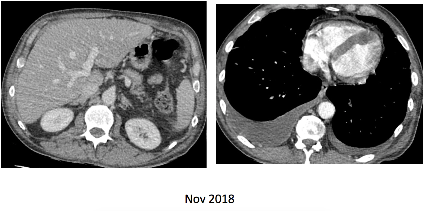
What would you report and would you add other images?
Update:
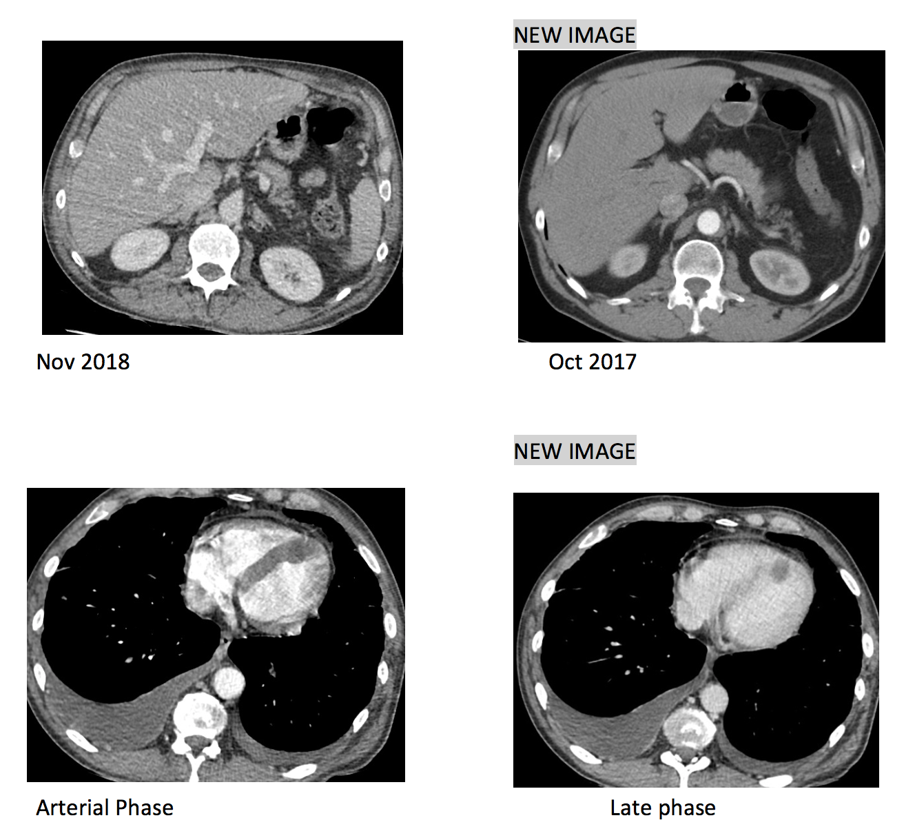
Ok friends, not many answers so far, so here are two additional images that may help you detect significant pathology. Do you see it now?
Click here for the answer
Dear friends: This is our last case for the season. I realize that the absence of a clear thoracic image probably made some people reluctant to give a chance to this case. ( Congratulations to those who answered).
But indeed, the chest is present in this case, and some findings were crucial for this patient
The abdominal CT shows some very small peritoneal nodules (red arrows) that, in the previous image one-year before were not present indicating, in an oncologic patient the presence of peritoneal metastases.

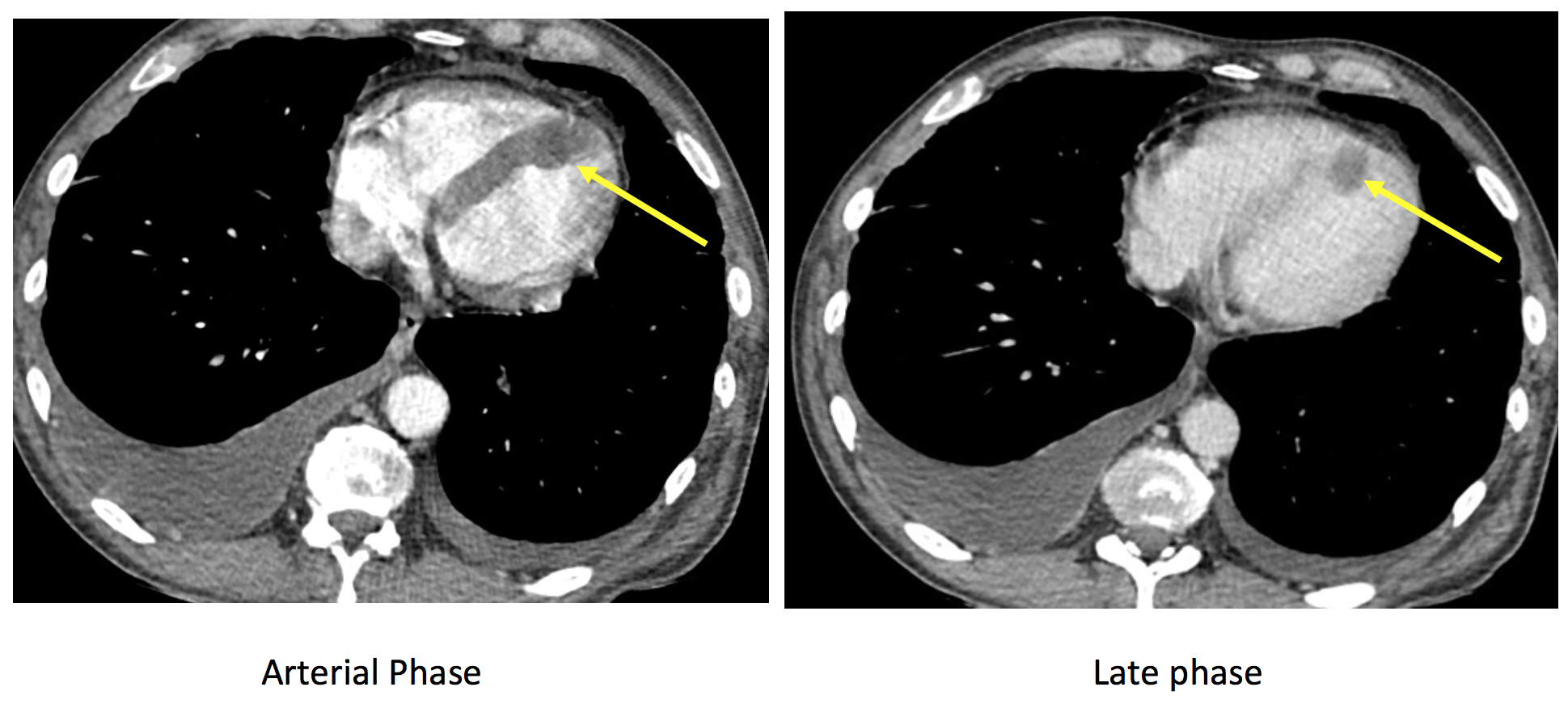
In the chest CT, the interventricular septum is thick and nodular (yellow arrow). In this case a late phase of the contrast (portal phase) allows a better delineation of a nodule and clearly defines a lesion. Again, being this a oncological patient with peritoneal metastases we must think that this, although less likely as a metastatic target, could be cardiac metastases.
Several main points to obtain from this case:
1- Always look at all the corners and edges of the images.
2- Compare with previous studies
3- Remember to look at the heart in Thoraco-abdominal oncologic CTs.
4- As Dr. Villanueva and colleagues from Surrey and Sussex, suggest that a late phase of the contrast, may help discover intracardiac metastases better. (Less density of the contrast as compared to the arterial phase).
5- Cardiac metastases are not so uncommon and account in some tumours up to 10% of the metastases. Look for them.
EPILOG:
Dear friends. This is our last case for the season. With the advent of the summer we will rest and try to enjoy as much as we can the nice weather, the company and the free time. I am aware that this has been a very strange and tragic year for many, but I also know that with this and other blogs we humbly contribute to maintain the flame of radiological knowledge. I have tried to present cases from many different origins, provided by friends from all over the world. My gratitude to all of them and to ESR and the help staff (Simona Bartovicova and Jabarkhel Kamil).
All our cases have had basics principles in common: to entertain, provoke and transmit some basic teaching points that may be useful in your radiological practice. As it is obvious, the chest, my preferred field in radiology, has dominated. Let me confess you that, after many years in thoracic radiology, I keep learning and finding new situations. The more I know the more I feel I need to know. It’s exactly the same feeling I have, as a veteran hiker when, after years wondering through my beloved Teruel mountains I discover new scenarios, new visions in a never-ending joy. Just wonderful.

1550 msasl. Virgen de La Vega. Alcalá de la Selva. Teruel. Spain. June 28 2009. J. Vilar
José Vilar


Metastasis at interventricular cardiac septum in apical segment?
Right adrenal and myocardial mets
bilateral pleural effusion rt > lt
deposit or thrombus in the myocardium
prpminent adrenals rt >lt
? ivc involvement
very small hypodense areas in rt lobe of liver inferior aspect
Right adrenal metastasis and interventricular metastasis