Dear friends,
This case goes to basics and it should not be difficult to interpret.
28-year-old. Incidental finding.
What finding is there and what do you think?
Check the images below!


Click here for the answer
Diagnosis: Pulmonary Sequestration
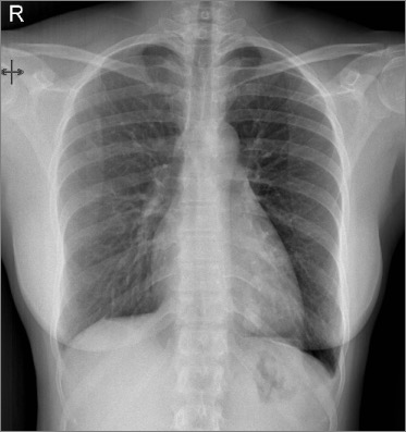
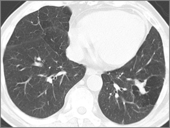
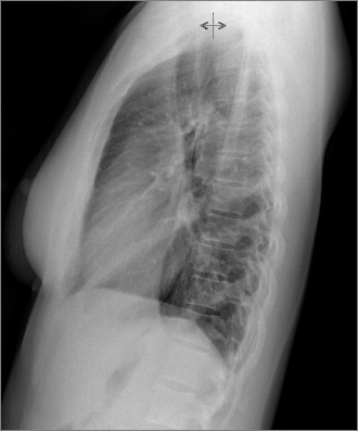
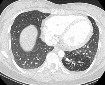
Comment: The plain films show an abnormal tubular opacity in the left lower lobe. There is some air trapping surrounding the lesion, better seen in in the CT (lung window).
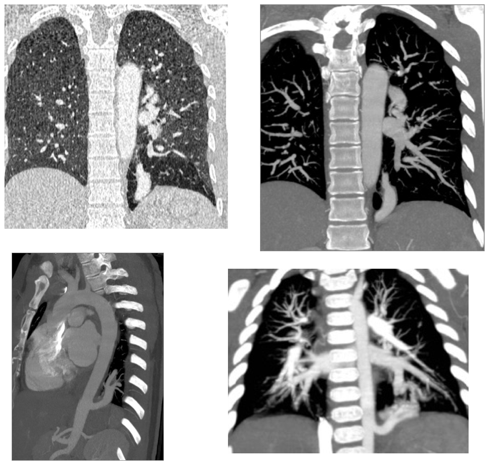
The CT angiogram reveals a large vessel connected to the descending aorta.
Intralobar pulmonary sequestration shares the visceral pleura with the normal lung usually occurs in the left lower lobe. The irrigation is systemic (aorta or branches) and the venous drainage to the pulmonary veins.
Teaching point: Air trapping is common in sequestration and can help you distinguish it from AV fistulae, but remember that tubular structures with air trapping also will occur with bronchial atresia or other causes of bronchial obstruction.
Bronchial atresia. Mucus filled bronchus simulates a vessel and air trapping.
Congratulations to those of you who mentioned the diagnosis, especially Trinity that did it immediately.


Good evening sir,
Chest X ray PA view shows a well defined oval shaped opacity in the left retrocardiac region.I am not sure about it’s calcification…but it does not show any cavitation or underlying bone erosion.No air fluid level seen within. Rest of the lungs are clear. Cardiac silhouette is normal. Bones and soft tissues also appear normal.
D/D is 1. SPN
2. Pulmonary Sequestration
3. Hamartoma if it’s calcification…
Regards.
Hi!
I agree with your differential diagnosis, I would like to add a pulmonary arteriovenous malformation, because I think that may exist a vascular dilated structure on top of the nodule that run to the hilium.
Regards!
Calcificaciones superpuestas mediastino, área retrocardiaca y hemidiafragma izquierdo (lineal). Signos de hiperinsuflación pulmonar. Enfermedad por exposición al asbesto.
There’s a tubular opacity in left lower lobe (retrocardiac). This structure seems to belong to pulmonary vessels. This finding is in a young assymptomatic patient, so I’d expect to find a pulmonary arteriovenous malformation (+/- congenital pulmonary airway malformation, due to the association).
OK, some of you are very good.! Sorry but we did not include the lateral in the first images. Here it comes and I am adding a lung window CT image and expect to see your comments..
Good afternoon sir.
The lateral chest radiograph shows some postero-inferior displacement of the oblique fissure and tubular oval opacity in retrocardiac region.
CT chest shoes obliteration of the brochure with distal hyperlucent lung,
Findings are suggestive of Bronchial atresia involving the LLL.
Regards.
Very good differential diagnosis. Tomorrow you will have the answer.
Tubular shadow in lll projection. Ct shows mucocele within hypoatenuating lung segment. Bronchial atresia, probabaly subsegmental bronchi of left lower lobe.
Had a case recently in a 45 year old, also asymptomatic, my seniors disagreed with me. But i stood my ground! 🙂