In the course of preparing a book on lung imaging that will be published to mark the International Day of Radiology (IDoR), the ESR spoke to Nicola Bedlington, executive director of the European Patients’ Forum (EPF), who shared her views on healthcare in the EU and explained why she chose to participate in IDoR 2013.
ESR: What is the overall aim of your organisation?
Nicola Bedlington: Our vision is high quality, patient-centred and equitable healthcare for all patients throughout the European Union.
The European Patients’ Forum is an umbrella organisation that works with patients’ groups in public health and health advocacy across Europe. Our members represent specific chronic disease groups at EU level, or are national coalitions of patients. We currently represent almost 60 such organisations.
Our mission is to be the collective patients’ voice at EU level, manifesting the solidarity, power and unity of the EU patients’ movement, and to provide a strong and united patients’ voice in order to put patients at the centre of EU health policy and programmes. In this regard we are the key interlocutor with EU institutions on cross-cutting issues affecting all patients.

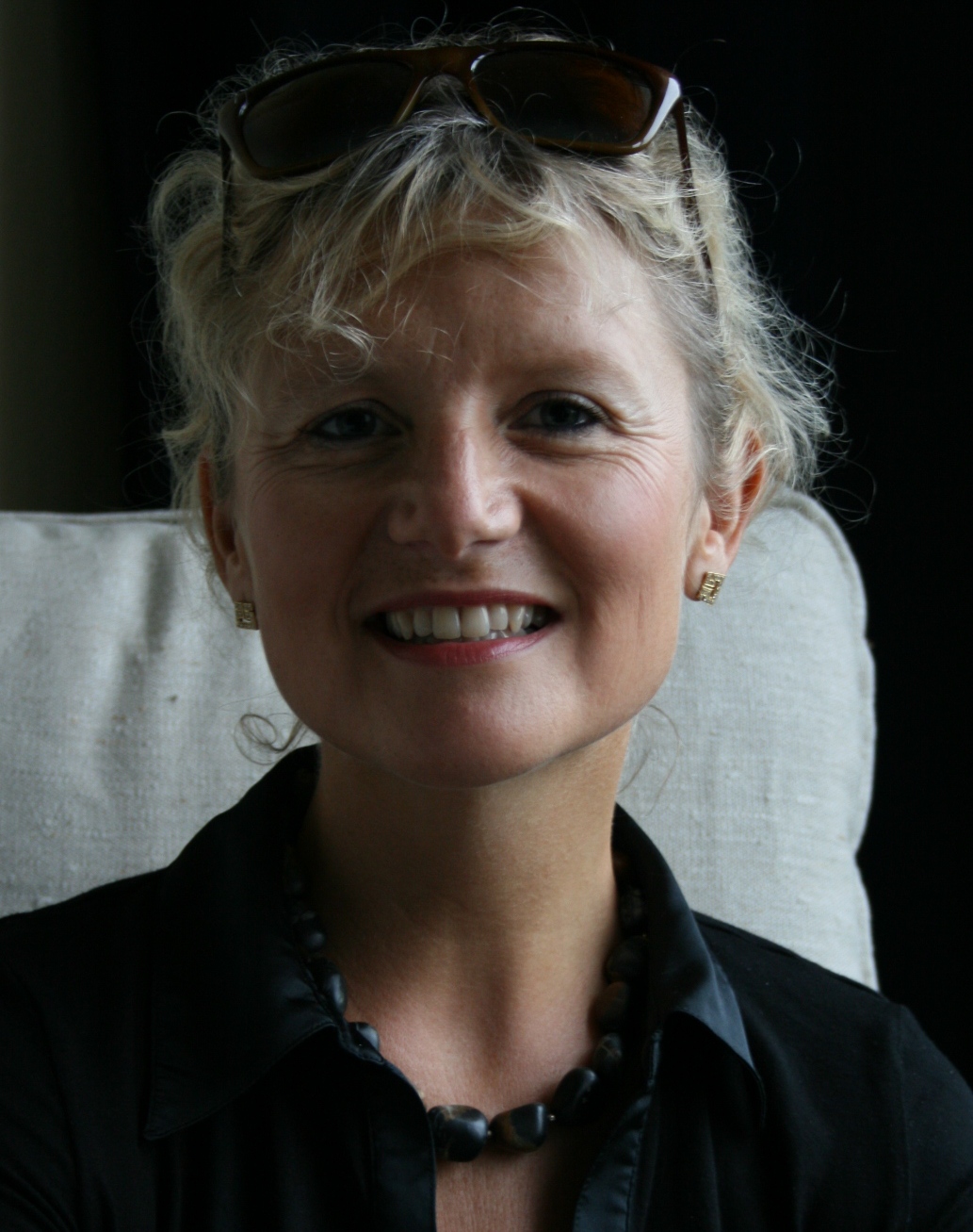
Nicola Bedlington, executive director of the European Patients’ Forum (EPF)
ESR: What exactly does your organisation do to meet this aim?
NB: The EPF helps to empower patients’ organisations through educational seminars, policy initiatives and projects. We coordinate best practice exchanges between patient organisations at European and national levels. Our programmes also help to strengthen organisational and advocacy capacity.
ESR: Your organisation has experience working with various chronic disease groups. Do many patients suffer from chronic diseases in the EU?
NB: Following consultation with our members we estimate there are at least 150 million patients with chronic conditions across the European Union. This figure is likely to increase given the ageing population.
ESR: Many EU countries face significant health budget cuts, leading to shorter hospital stays and less access to modern equipment (i.e. long waiting lists for MRI exams). How can patient care be promoted in this context?
NB: The EPF is working with its member organisations to ensure health is seen as an investment, and patients are not perceived as purely cost drivers. Major health inequalities exist across the EU which impact enormously on patients’ access to care.
Building on the three pillars of quality information, health literacy and empowerment, patients can be agents of change and sources of innovation, particularly in terms of equity and sustainability of care. There need to be meaningful opportunities for patient involvement throughout the healthcare sector. We promote meaningful patient involvement in all forms of innovation, whether it is in high or low technology, pharmaceuticals, information technology, social change or systems change. The patient community seeks partnerships with researchers, policy-makers and industry in order to achieve greater impact in this arena.
Read more…