Muppet cannot find difficult cases and, despite himself, is forced to show easy ones. Today we’re showing radiographs of a 48-year-old woman who has had moderate dyspnea for a while.
Diagnosis:
1. Swyer-James syndrome
2. Tumour left main bronchus
3. Old TB left lung
4. None of the above
This week I’m presenting you another new ‘Face the Examiner’ case which simulates a real examination. Showing radiographs of a 35-year-old male with high fever and left pleuritic pain.
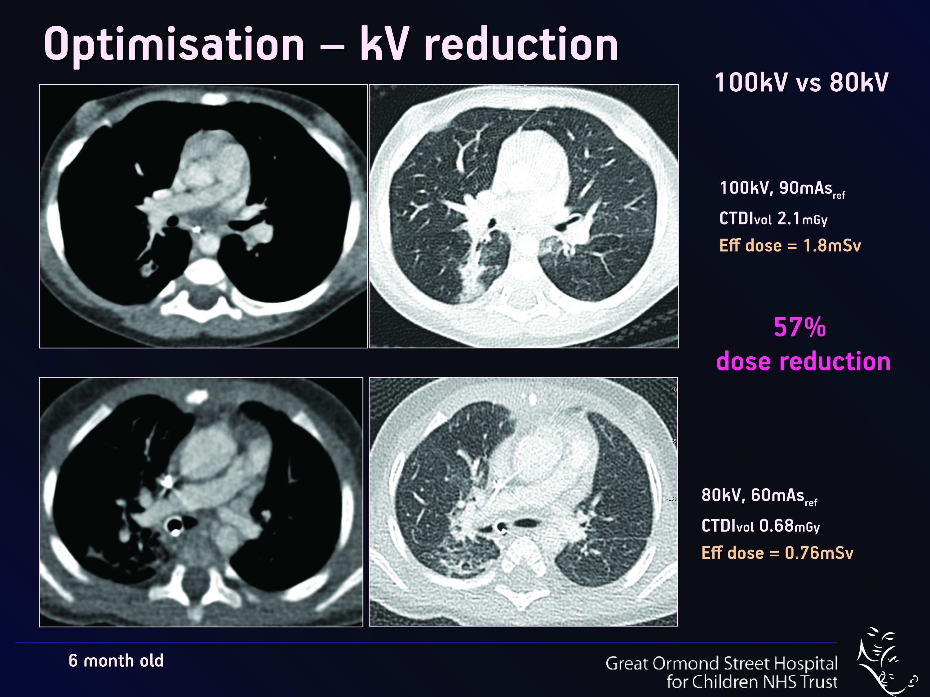
The optimisation and justification of procedures is vital when using CT as an imaging modality; particularly on children, who are more sensitive to ionising radiation than adults. Therefore, it is crucial that all those who use CT understand the physics behind the equipment and ultimately use this understanding to minimise the potential risks while maximising the potential benefits to each individual patient. Patients should also be informed of the risks and benefits of undergoing a CT scan. World-renowned experts will explain these issues in detail during a Special Focus Session at ECR 2013.
“Not all radiologists and technicians are aware of the latest dose reduction strategies. Some are not necessarily so well-informed and perhaps do not realise how important this is. We believe that it is a question of trying to get everybody to a certain level of knowledge and expertise,” said Dr. Catherine Owens, paediatric radiologist and CT unit lead at Great Ormond Street Children’s Hospital in London, U.K.
Heart disease affects a very large number of people worldwide, and the consequences can be serious and even lethal. Here, and perhaps more than in many other areas of medicine, imaging has helped to improve treatment and prevention. It does so by detecting the disease at an early stage, sometimes even before its emergence, especially in patients at risk of ischaemic heart disease.
Today, diagnosing cardiac patients has become routine for many radiologists. However, some of them may not know of recent developments in this field and they may need to refresh their knowledge. A panel of experts will update both general and specialised radiologists with the latest information available on cardiac imaging, during the dedicated Mini Course ‘Organs from A to Z: Heart’ at ECR 2013. After an introduction to heart anatomy and the main imaging protocols, the course will focus on valvular diseases and cardiomyopathies; two pathologies commonly encountered in radiology practices.
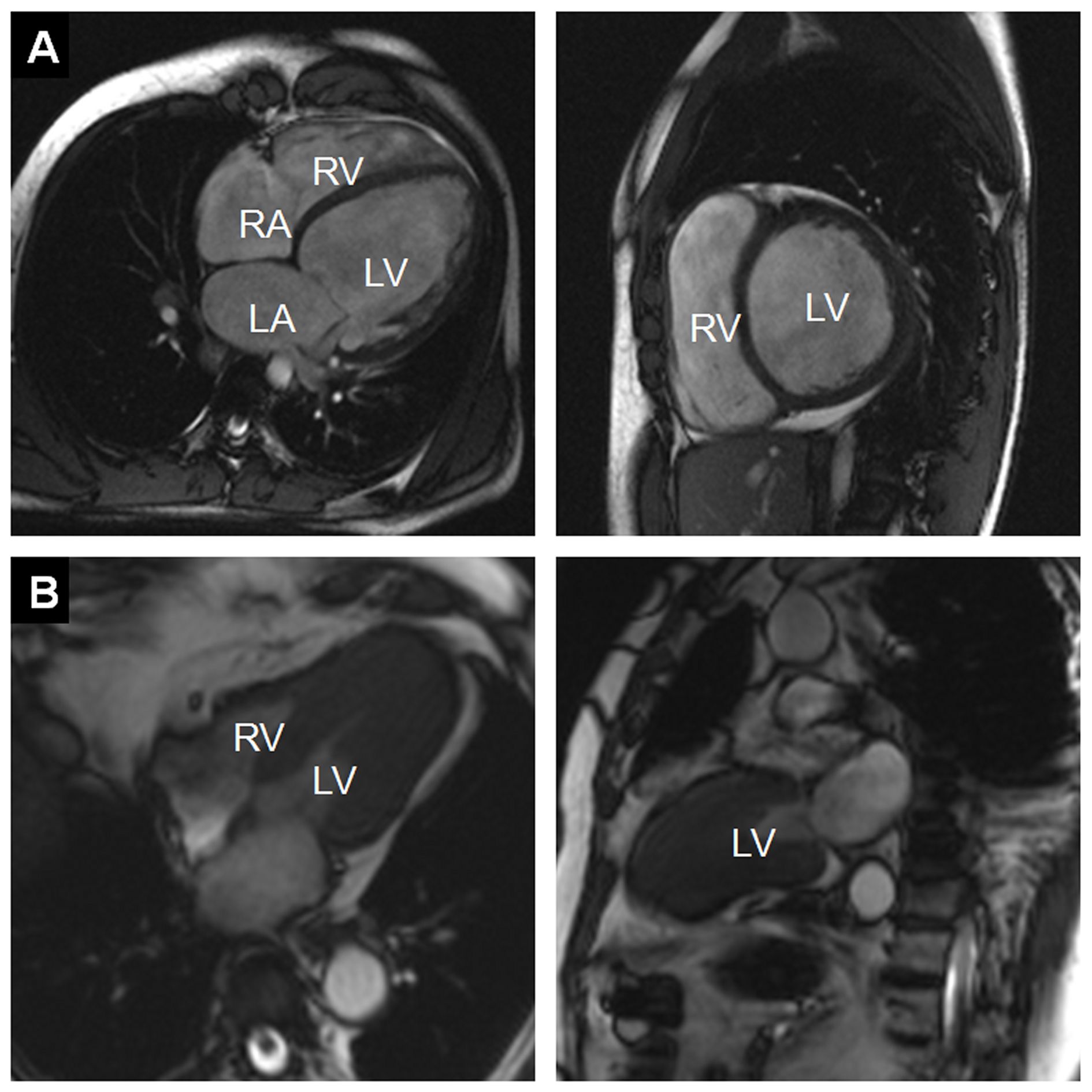
Figure 1: A) Example of a dilated cardiomyopathy (DCM). Cine-MR images in four-chamber view (left) and short-axis view (right) at end-diastole show significant dilatation of the LV cavity. Ejection fraction was <35% in this patient. (RA = right atrium; LA = left atrium; RV = right ventricle; LV = left ventricle) B) Example of an asymmetrical, apical hypertrophic cardiomyopathy (HCM). Cine-MR images in a four-chamber (left) and two-chamber view (right) in systole show a markedly thickened left ventricular myocardium predominantly of the apex, as compared with the basal segments (RV = right ventricle; LV = left ventricle).
The brain remains undoubtedly one of the most mysterious organs of the human body. Magnetic resonance imaging has helped to unveil some of its secrets, and major advances have been made in understanding how the brain functions. Recent developments with resting fMRI (rfMRI) and diffusion MRI (dMRI) indicate that scientists are beginning to see beyond the brain: they have actually started to visualise the human mind. This new information is particularly relevant for understanding complex processes such as dementia, autism and depression. It is also proving increasingly central to the diagnosis of comas and chronic disorders of consciousness.
Leading researchers will discuss where the latest advances have led them and what the future will bring in a dedicated New Horizons Session during ECR 2013. FMRI has been used for over twenty years to visualise changes in brain activity by comparing a task versus a control task, and showing and quantifying how much brain activity is involved in the process. The recent addition of rfMRI enables researchers to track networks that are randomly active. A patient lying in a scanner with no particular task to perform will usually start thinking about the trivialities of the day and go from one thought to the other (“Did I close the door before I left? What am I doing here?” etc.). Neuroresearchers can track this mind mumbling with complex mathematics and extract information from what they call the default mode network.
Fig. 1: Differences in functional connectivity from rfMRI between autistic patients and age- and gender matched controls: the major disconnection is between the cerebellum and frontal language areas.
The quality of the ECR’s sessions for radiographers has been given a welcome seal of approval from the European Federation of Radiographer Societies (EFRS) who recently elected the ECR as their official annual scientific meeting. EFRS president, Prof. Graciano Paulo, from the college of health technology of Coimbra, Portugal, has been coming to the ECR for more than a decade and firmly believes the upcoming congress boasts one of the best selections yet for radiographers. Here he gives his personal preview of ECR 2013 and each of these sessions, all of which you can find in the ECR 2013 Interactive Programme Planner by searching for ‘radiographers’.
Read on for Prof. Paulo’s preview of all the sessions for radiographers at ECR 2013 …
As some of you may be taking the Diploma examination during ECR 2013, I’d like to introduce a new type of post that I call ‘Face the Examiner’. Its purpose is to simulate a real examination: images will be shown and you will be asked to describe the findings. You should then offer a differential diagnosis and suggest a procedure that will confirm your preferred option.
The only difference with respect to a real examination is that you will be given the correct answers during the exercise. I’ll try to keep it simple by not giving long lists of possible diagnoses and by making sure the possibilities are coherent with the imaging features.
Our first ‘Face the Examiner’ case concerns preoperative chest radiographs in a 75-year-old man with prostate carcinoma.