José Vilar and Friends Case 7 (Update: Solution!)

Good day colleagues,

This case was brought to me by Drs María Vega, Magdalena Graells and Luis Garcia (Hospital Universitario Dr Peset, Valencia; all pictured above in the case cover) and opens a new look to my blog.

This is a 35-year-old man from Pakistan that complains of long standing back pain.

Read more…

01
Apr 2019
POSTED BY
DISCUSSION 11 Comments

ECR 2019 Cases of the Day Quiz Winners

The Cases of the Day Quiz featured 20 challenging cases covering different sections of radiology.
The cases were shown during ECR 2019 and attendees were encouraged to submit their diagnosis.
From the 374 participants, the following solved most cases correctly:

Elliott Rees; Sawston, Cambs/UK
Filip Vanhoenacker; Antwerp/BE
Ümit Tüzün; Istanbul/TR
Artur Komorowski; Owczary/PL
Manabu Minami; Yokohama/JP
Jan Balak; Prague/CZ
Martina Slamova; Nove Mesto Na Morave/CZ
Diogo Roriz; Coimbra/PT
Veronika Pesti; Budapest/HU
Timothy Sadler; Cambridge/UK
Miguel Correia da Silva; Porto/PT
Vendoti Nitheesha Reddy; Bengaluru/IN

To view the cases of ECR 2019 please click here. 

Congratulations to all winners!

20
Mar 2019
POSTED BY
POSTED IN ,
DISCUSSION 0 Comments

The best submissions for the “Just a cyst?” interlude at ECR 2019

Dear Friends,

Over the last couple of years, one of the last sessions at the ECR has always covered 20 interesting cases from various subspecialties, which the audience is asked to solve in an interactive way to broaden and update their knowledge.

In between, the very best submissions from the global radiological community have been presented in an interlude lecture. The best submission has always been awarded with a prize and a certificate.

Due to time limits, not all submitted cases can actually be shown onsite, but the session’s rising popularity has resulted in increasing numbers of submissions of excellent quality and didactic value. This is why we would like to give our submitters the opportunity to reach a broader audience by posting the best cases here on the ESR Blog.

We all use the term “cyst” a great many times every day. It comes in handy, when we are dealing with something roundish and not solid. Mostly, we want to convey the message to clinician and patient: “nothing to worry about, just fluid”. But is it always that simple? What does the location tell us and are there any additional findings useful to report? Are they “leave me alone” lesions by definition or should we sharpen our awareness to separate the needle in the haystack? When you start to think about it, you might run into the risk of seeing danger everywhere.

Below, we are pleased to present the best submissions for the “Just a cyst” interlude at ECR 2019:

19
Mar 2019
POSTED BY
POSTED IN
DISCUSSION 0 Comments

José Vilar and Friends Case 6 (Update: Solution!)

Hello friends,

this time we are going to move down into the abdomen. This case was shown to me by my good friend Dr José Vizuete (Hospital Universitario Dr Peset, Valencia) and I think that it shows how we should look at images and use all the information included in them.

This is a 39-year-old woman who presents at the Emergency Room with abdominal pain in two occasions.
Read more…

18
Mar 2019
POSTED BY
DISCUSSION 4 Comments

José Vilar and Friends Case 5 (Update: Solution!)

Dear friends,

Today’s radiographs belong to a 37-year-old with fever and cough. Rule out pneumonia.
Read more…

04
Mar 2019
POSTED BY
DISCUSSION 9 Comments

Incoming ESR President lays out clear vision for ECR 2020

By Julia Patuzzi

It is a well-established tradition that on the final day of the congress, ECR Today looks ahead to next year’s ECR. We therefore spoke with Professor Boris Brkljačić from Zagreb, Croatia, the incoming ESR President, who is in charge of ECR 2020. He shared with us some of his ideas and plans for the next European Congress of Radiology.

ECR Today: Professor Brkljačić, the first visual impression of the next congress is always the congress poster. For ECR 2020 you chose artwork by the award-winning Canadian illustrator Peter Diamond, depicting a young woman looking at a small object floating just above her cupped hand. Can you tell us a little about how this particular design came about?

Incoming ESR President Boris Brkljačić is professor of radiology and vice-dean at University of Zagreb School of Medicine, Zagreb, Croatia, and chair of the Department of Diagnostic and Interventional Radiology of University Hospital ‘Dubrava’ in Zagreb.

Boris Brkljačić: The ESR Office provides several options for the congress poster, created by professional designers, and the Congress President and PPC members select one. The selected solution was the best among the proposed options. It resembles Rembrandt’s artwork, with sharp light and dark contrast, and is in good accordance with the slogan for ECR 2020: ‘A Clear Vision for Radiology’. The small floating object represents artificial intelligence, which will be an important topic at the congress, and the names of the ECR 2020 ‘ESR meets’ countries are visible at the bottom of the poster. The 2020 poster contains fewer colours and illustrations compared to the 2019 poster, and is concordant with the visual style of the ESR’s main scientific publication, the journal European Radiology.

ECRT: As the new ESR President, you are also chairperson of the Programme Planning Committee for ECR 2020, which has already been working on preparing the scientific programme for a few months. Can you tell us something about the highlights of the 2020 programme or any specific focus we can expect?

BB: I am very fortunate to have selected excellent Programme Planning Committee members, who are hard-working and dedicated experts in their fields. Planning has already been running at full speed for a few months in order to create a well-balanced programme of very high-quality professional, educational and scientific content. New Horizons Sessions, State of the Art Symposia and Special Focus Sessions have already been selected and mostly created; they are very relevant and balanced, so that young radiologists and experts in particular radiological fields will have interesting sessions to choose from in all areas of radiology. Emerging and hot topics will be covered, like lung cancer screening, artificial intelligence, stroke diagnosis and treatment, and many others. I expect that the plenary/honorary sessions should be the highlight of the congress, as they are interesting for all participants, regardless of their age and expertise. Read more…

How to deal with common diseases in Pakistan: the radiologist’s perspective

by Katharina Miedzinska

Each country has its own special healthcare challenges to shoulder. In Pakistan, a densely populated country located in South Asia, with an estimated population of more than 200 million, major healthcare challenges include exceptionally high prevalence rates for certain diseases. During today’s ‘ESR meets Pakistan’ session, some of the country’s top radiologists will discuss the role radiology plays in managing three of the most common ones: oral cancer, chronic hepatitis and tuberculosis.

Many diseases are common in Pakistan, among them endemic and epidemic infectious diseases, emerging infections, and an increasing burden of non-communicable diseases. The actual burden of infections with the hepatitis B and C virus (HBV, HCV) is approximately seven percent, making Pakistan a country with one of the highest prevalence rates for viral hepatitis in the world. Approximately ten million people in Pakistan are infected with HCV alone. Following the acquisition of the virus, acute HCV infection can progress to chronic infection, which in turn is associated with several morbidities, such as liver cirrhosis and cancer. Besides hepatitis, also malaria, the polio virus (which still circulates in core-reservoirs across Pakistan, although with lesser intensity), dengue outbreaks, and other infectious diseases pose a serious threat to public health security. Over the past few decades, Pakistan has suffered a great deal from these and other infectious diseases. Global warming, changing climate conditions, environmental degradation, and other ecological determinants have a direct effect on these diseases and result in the emergence or re-emergence of infectious entities. The causes of such disease outbreaks are complex and often not well understood.

Read more…

03
Mar 2019
POSTED BY
POSTED IN , ,
DISCUSSION 0 Comments

CEUS ready for clinical paediatric use argues UK expert

Professor Paul Sidhu is Professor of Imaging Sciences at King’s College London and consultant radiologist at King’s College Hospital, where he helped pioneer contrast-enhanced ultrasound (CEUS), a technique that can potentially be used to image children just as well as adults, in reduced time, and without the downsides of radiation and toxicity. Professor Sidhu will make the case for CEUS use in paediatric radiology today during the Luigi Oliva Honorary Lecture.

There is no question that ultrasound is the most child-friendly imaging technique. The modality has countless benefits for young patients: no ionising radiation, no sedation or anaesthesia, and ease of use, to name a few. “Ultrasound is the most suitable modality for imaging children, as they don’t have to stay still. Parents can be present during the examination to support their child. The radiologist can stop and start the examination without losing information, plus children have low body fat and are better suited to ultrasound,” Sidhu said.

Professor Paul Sidhu from King’s College London will argue for CEUS use in paediatric radiology in today’s Luigi Oliva Honorary Lecture.

Adding a contrast agent to ultrasound has been shown to improve diagnosis tremendously in applications where vascularity or haemo-dynamics must be assessed in real time. With CEUS, in the case of focal liver lesions, which are often difficult to characterise in adults and children on B-mode ultrasound alone, essential information from the arterial and portal venous phases aids diagnosis in minutes without needing CT or MR imaging. This alone should be a decisive argument for widespread CEUS use in the paediatric population, Sidhu explained.

“The combination of contrast with ultrasound allows for a more detailed assessment of the pattern of vascularity and dynamics in a continuous fashion for three to five minutes in real time, and it can be repeated as many times as necessary. The child does not need to keep still, and all the same focal liver characteristics seen in the adult are seen in the child. The ability to come to a clear diagnosis, with a single imaging examination is a great relief for the parents, often present with the child during the examination, rather than remote as with CT or MR imaging,” he said.

With CEUS, radiologists can obtain a lot of information in the first five minutes following the injection, the time window that the contrast agent lasts in the blood pool. This is more than enough to carry out the examination and make a conclusion. The technique can be used in every possible scenario from trauma to disease imaging, with great results. “Imaging with ultrasound in children is the most useful and informative procedure,” Sidhu concluded. Read more…

African radiologists call for more cooperation with the ESR in radiation protection

African radiologists will share their knowledge and experience of medical imaging practice in their respective countries, today during the ‘ESR meets Africa’ session. Examples of cooperation with the European Society of Radiology will be presented, notably in radiation protection, a field where efforts between Europe and Africa are starting to pay off.

Africa is big, diverse and full of possibilities. Its myriad of countries (54), languages, cultures and economic scenarios offers unmatched potential, but it can also complicate the organisation of radiology.

Prof. Dina Husseiny Salama and Prof. Hassen Gharbi, together with ESR President Prof. Lorenzo E. Derchi and Prof. Guy Frija, Chair of EuroSafe Imaging, surrounded by delegates at the 5th African Society of Radiology conference in January 2019, in Cairo, Egypt.

Equipment and workforce tend to vary considerably from one country to another. For example, the radiologists’ ratio ranges from 1 to 80 per million population, depending on the country.

“Our main challenges are in training and the implementation of national legislation that can help organise the field, especially regarding radiation protection,” said Prof. Hassen Gharbi from Tunis, Tunisia, who will co-chair the session with ESR President Lorenzo Derchi from Genoa, Italy.

Technological advances have opened new horizons for the application of ionising radiation in healthcare all around the world and this has led to an increase in medical imaging procedures using radiation, also in Africa.

Unlike most of the continent, Tunisia, Algeria and Morocco have dedicated guidelines on ionising radiation use. But even there, rules must be implemented to help healthcare professionals prescribe examinations adequately.

“There is a growing need for structured strategies and a holistic approach towards the full integration of radiation safety and clinical imaging guidelines in Africa,” said Prof. Dina Husseiny Salama from Cairo, Egypt. Read more…

Systemic effects in interventional oncology: Holy Grail or Pandora’s Box?

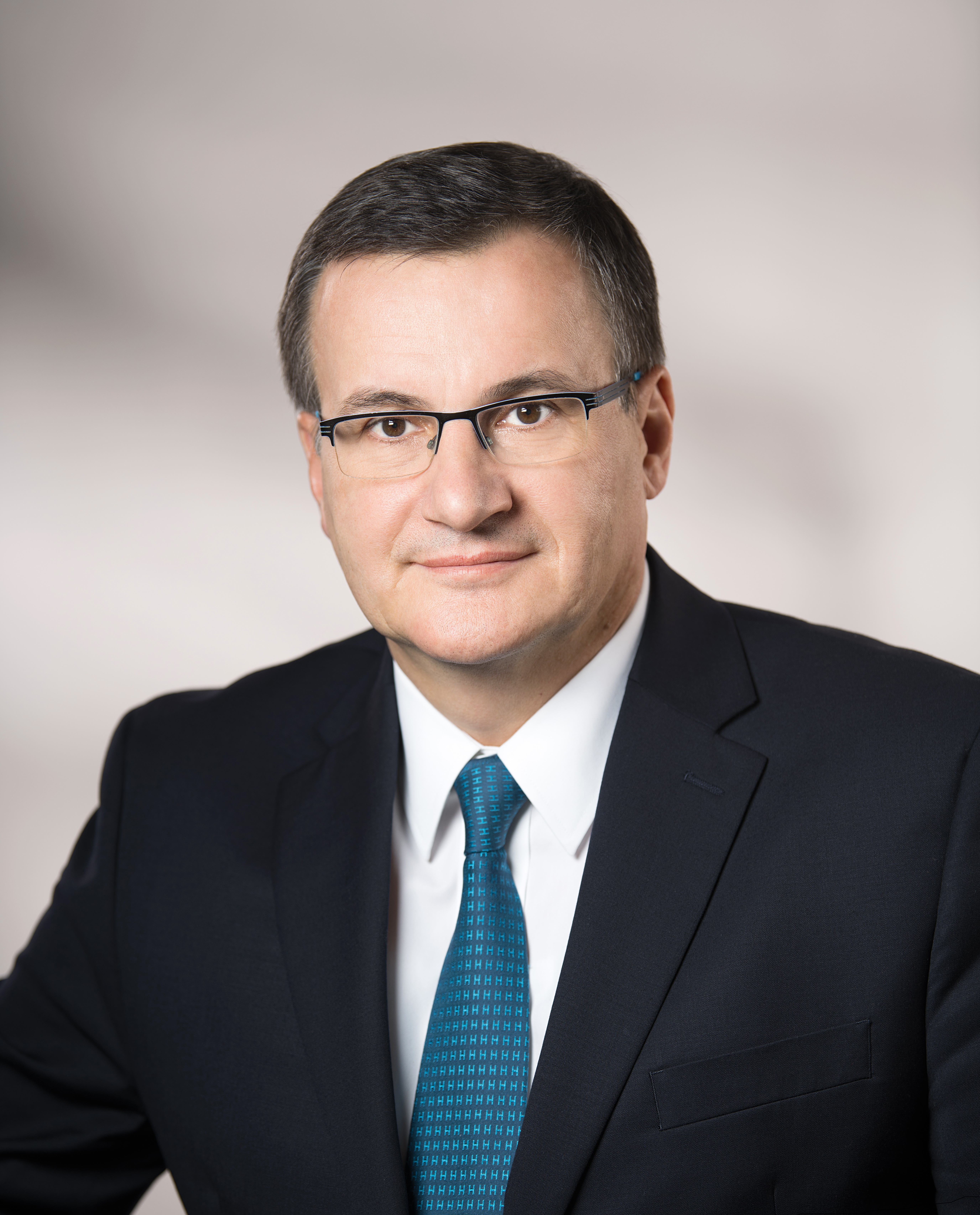
Nahum Goldberg is full professor of radiology at Hadassah Hebrew University, Jerusalem, Israel and visiting professor of radiology at Harvard Medical School, Boston, MA. He is a worldwide expert in image-guided tumour therapy, a field he has helped pioneer and continues to advance. He will share some of the latest results on the effects of these therapies, both positive and negative, today during the Josef Lissner Honorary Lecture.

Image-guided therapy and transcatheter intervention work best on small tumours in the liver, kidney and several other organs, evidence over the past two decades has showed. That was the birth of interventional oncology and it held exciting promise for the (radical) improvement of cancer treatment. Based on that knowledge and promise, many researchers have worked towards combining these procedures with more conventional cancer therapies like chemotherapy and radiation to increase the size and range of the population that can be treated.

Prof. S. Nahum Goldberg from Jerusalem will share some of the latest results on the effects of image-guided tumour therapy, during the Honorary Lecture today at 12:15.

One of the underlined premises of interventional therapies is that using imaging to guide a procedure is less invasive than using other methods like surgery. Despite this and other advantages that have led to ablation systems and techniques now being used clinically more than 100,000 times annually, things have not always gone as planned, according to Goldberg, who heads the interventional oncology unit at Hadassah Medical Centre.

“Specifically, for much of the last two decades, we have argued that other benefits of these interventional oncologic therapies, including percutaneous tumour ablation and chemoembolisation, were focal and local, based upon our conviction that we were only affecting the tumour we were treating and not the entire patient system. It turns out, based on early case reports and more solid evidence produced over the last five years or so, that this premise is not always true,” said Goldberg.

“Most of the damage occurs where intended, but all kinds of pathways in the system are also activated after an intervention. Just like a sunburn that affects a local piece of skin can be accompanied by a headache or fever, a systemic reaction can follow an interventional oncologic procedure in some patients and under certain circumstances. Depending on the type of tumour and its location, a series of both positive and negative systemic effects can be unleashed,” he added.

These effects are rather variable and are currently hard to predict, but one thing is certain: there is a good side and a bad side – the Holy Grail and Pandora’s Box, as Goldberg put it. Read more…