Dear Friends,
The fifth chapter of “The wisdom of Dr. Pepe” is the last presentation of this blog series and ends the present season.
The axial CT images below belong to a 37-year-old woman, who was operated on five years ago for a retroperitoneal tumour. What do you see?
Check the images below, leave your thoughts in the comments section, and come back on Friday for the answer.
Read more…
Dear Friends,
Today we are showing a case in recognition of the International Day of Radiology, which takes place tomorrow.
Below are images of a 35-year-old woman with chest pain and progressive dyspnoea for the last three weeks. Leave your thoughts in the comments section and come back on Friday for the answer.
Most likely diagnosis:
1. Lymphoma
2. Pleural metastases
3. Mesothelioma
4. Any of the above
Read more…

Dear Friends,
Welcome back! To start the academic year I would like to show a case seen by Dr. Pepe while I was vacationing in Minorca.
Radiographs belong to a 35-year-old woman with pain in the mandible for the last two years. CT of the mandible before biopsy and pre-op PA chest radiograph are shown. After seeing both, Dr Pepe suggested a diagnosis. What do you think?
Check the images below, leave your thoughts in the comments section, and come back on Friday for the answer.
Read more…

Dear Friends,
Muppet and I are very happy to have reached one hundred cases. We hope you enjoyed them as much as we did. Radiographs of this case belong to a 52-year-old man with vague chest complaints. He was operated on for testicular tumour fifteen years earlier.
Check the images below, leave your thoughts and diagnosis in the comments section, and come back on Friday to find out the answer.
Diagnosis:
1. Duplication cyst
2. Lymphangioma
3. Metastasis from testicular tumour
4. None of the above
Read more…
Dear Friends,
Muppet wants to start the new season with a warm-up case, provided by my good friend and former resident Carles Vilá. Images belong to a 62-year-old woman who has had a dry cough for the last six months. Chest radiograph was normal and a CT was taken.
Diagnosis:
1. Coin
2. Chicken bone
3. Pencil lead
4. None of the above
Read more…

Dear Friends,
Dr. Pepe has urgent business (suspect he is visiting Miss Piggy) and asked me to cover for him this week and the next. He will be back with a new Diploma case on Monday, June 9.
My good friend and former resident Dr. Eva Castañer has contributed with this week`s case. Images are of a 74-year-old male admitted with hemoptysis. Leave your diagnosis in the comments section and come back for the answer on Friday.
Diagnosis:
1. Pulmonary infarction
2. Carcinoma
3. Tuberculoma
4. None of the above
Read more…

Dear Friends,
I am back with radiographs of a 63-year-old woman with malaise and low-grade fever. Check the images below and leave me your thoughts and diagnosis in the comments section. Come back on Friday for the answer.
Diagnosis:
1. Carcinoma of the lung
2. Pneumonia
3. Thymoma
4. None of the above
Read more…

Dear Friends,
This week we have the case of a 45-year-old man, who is an alcoholic with abdominal pain, jaundice, and weight loss.
Possible diagnoses:
1. Duodenal neoplasm
2. Focal pancreatitis of pancreatic head
3. Pancreatic neoplasm
4. None of the above
Read more…

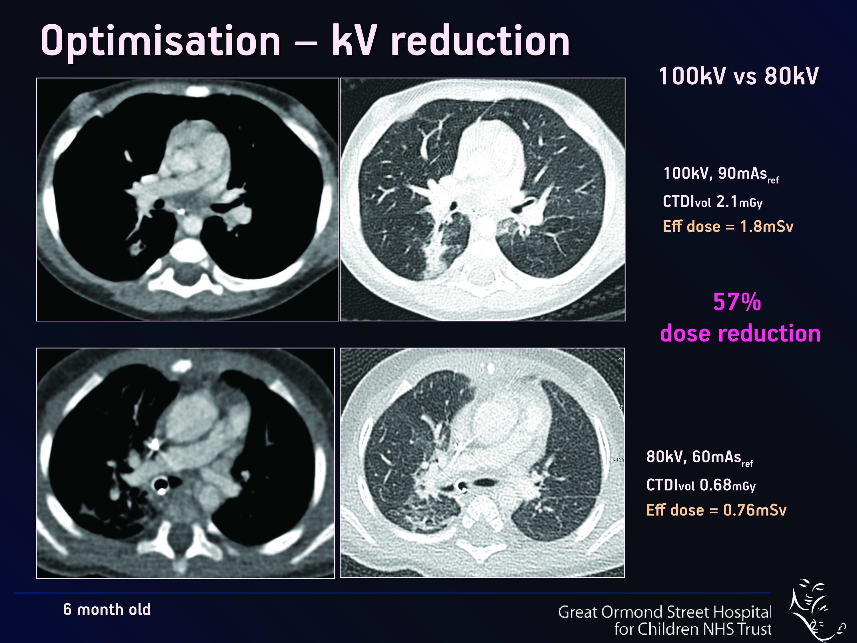
The optimisation and justification of procedures is vital when using CT as an imaging modality; particularly on children, who are more sensitive to ionising radiation than adults. Therefore, it is crucial that all those who use CT understand the physics behind the equipment and ultimately use this understanding to minimise the potential risks while maximising the potential benefits to each individual patient. Patients should also be informed of the risks and benefits of undergoing a CT scan. World-renowned experts will explain these issues in detail during a Special Focus Session at ECR 2013.
“Not all radiologists and technicians are aware of the latest dose reduction strategies. Some are not necessarily so well-informed and perhaps do not realise how important this is. We believe that it is a question of trying to get everybody to a certain level of knowledge and expertise,” said Dr. Catherine Owens, paediatric radiologist and CT unit lead at Great Ormond Street Children’s Hospital in London, U.K.
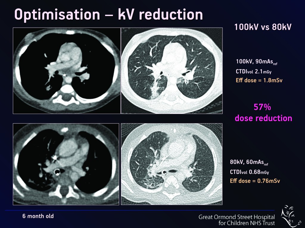
Fig. 1
Read more…

Dear Friends,
Muppet wishes to present the case of a 75-year-old woman with bilateral mastectomies for carcinoma 10 and 7 years previously. Chest radiographs and CT are shown.
Diagnosis:
1. Pleural metastases
2. Mesothelioma
3. Pleural TB
4. None of the above
Read more…