ECR 2013 Focus: Organs From A to Z: Heart

Heart disease affects a very large number of people worldwide, and the consequences can be serious and even lethal. Here, and perhaps more than in many other areas of medicine, imaging has helped to improve treatment and prevention. It does so by detecting the disease at an early stage, sometimes even before its emergence, especially in patients at risk of ischaemic heart disease.
Today, diagnosing cardiac patients has become routine for many radiologists. However, some of them may not know of recent developments in this field and they may need to refresh their knowledge. A panel of experts will update both general and specialised radiologists with the latest information available on cardiac imaging, during the dedicated Mini Course ‘Organs from A to Z: Heart’ at ECR 2013. After an introduction to heart anatomy and the main imaging protocols, the course will focus on valvular diseases and cardiomyopathies; two pathologies commonly encountered in radiology practices.
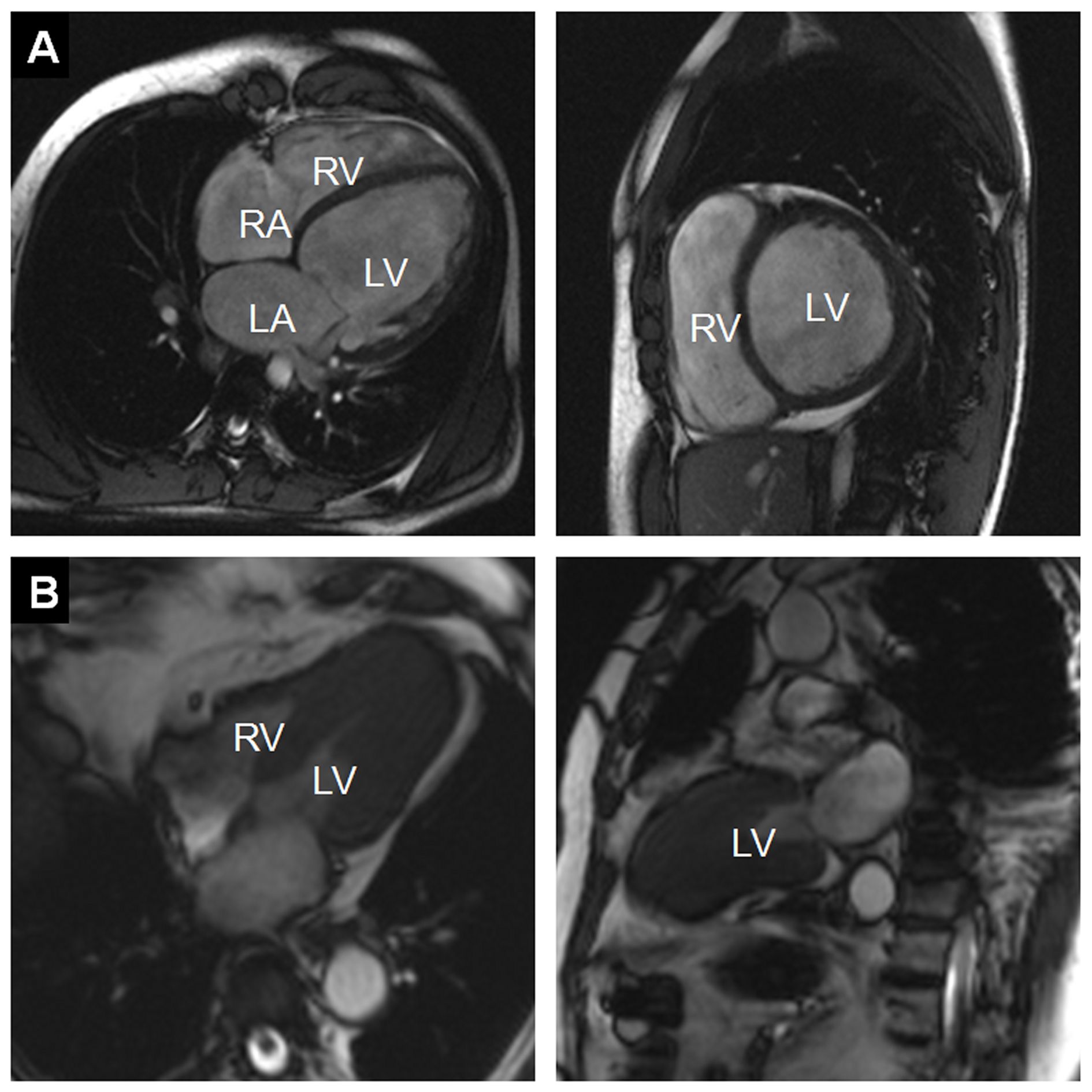
Figure 1:
A) Example of a dilated cardiomyopathy (DCM). Cine-MR images in four-chamber view (left) and short-axis view (right) at end-diastole show significant dilatation of the LV cavity. Ejection fraction was <35% in this patient. (RA = right atrium; LA = left atrium; RV = right ventricle; LV = left ventricle)
B) Example of an asymmetrical, apical hypertrophic cardiomyopathy (HCM). Cine-MR images in a four-chamber (left) and two-chamber view (right) in systole show a markedly thickened left ventricular myocardium predominantly of the apex, as compared with the basal segments (RV = right ventricle; LV = left ventricle).