Dr. Pepe’s Diploma Casebook: Case 10 – SOLVED!

Dear Friends,
This week’s case is an 85-year-old man with weight loss. Would you call this study normal?
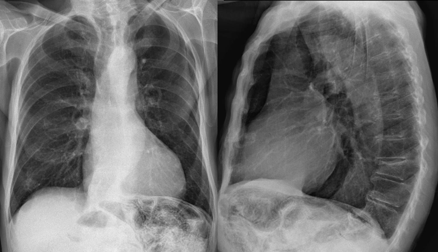
Fig. 1: 85-year-old man with weight loss
Findings:
Diffuse, curvilinear, low-density lines (arrows) in the small bowel suggest intramural gas collections.
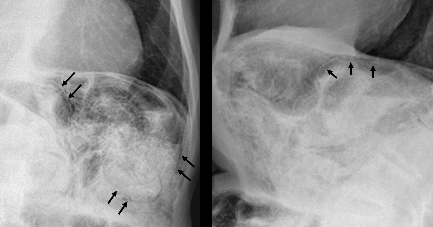
Fig. 2
CT confirms air in the jejunal walls (black arrows). No free air is seen.
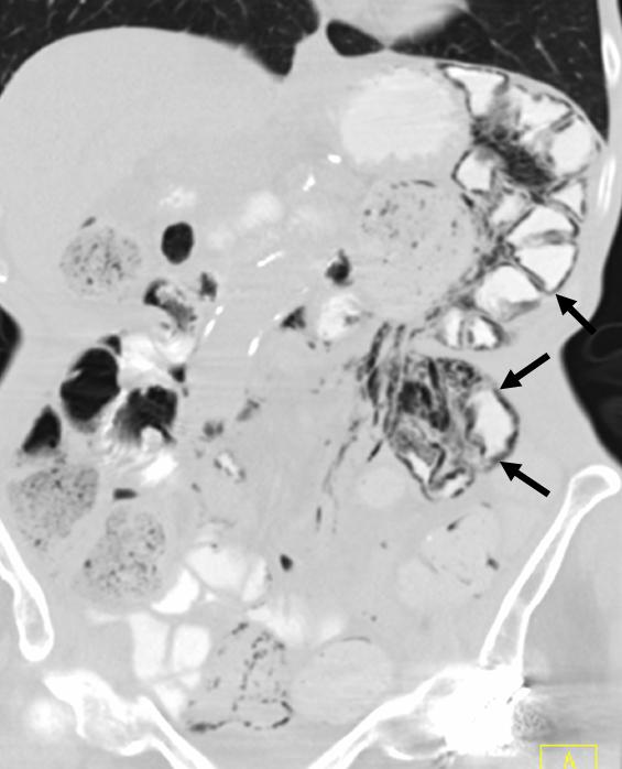
Fig. 3
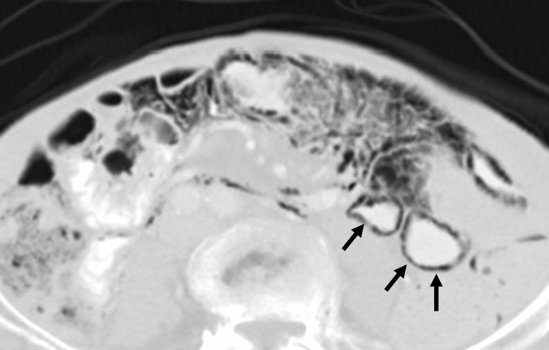
Fig. 4
Final answer: Pneumatosis intestinalis, secondary to bowel ischemia
Pneumatosis intestinalis (PI) is defined as the presence of gas in the bowel wall.
On both radiography and CT, PI usually appears as a low-density linear (Fig. 5a, arrows) or bubbly (Fig. 5b, arrows) pattern of gas. PI is a sign, not a disease, and it must be interpreted relative to the patient’s overall clinical condition.
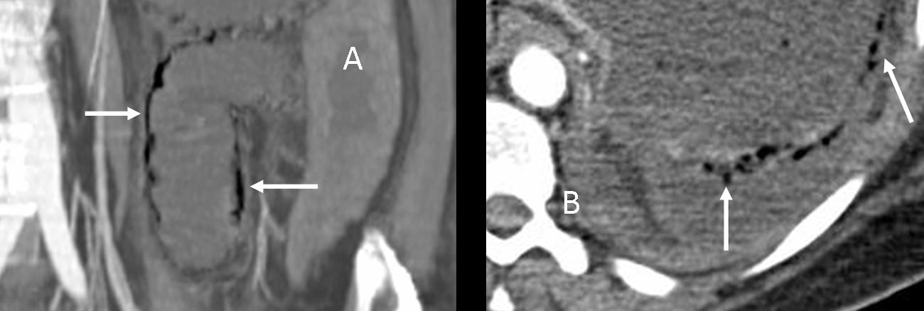
Fig. 5
PI has many causes, ranging from benign to life-threatening conditions. Clinical symptoms and laboratory data provide important clues to clarify whether PI is due to a benign or life-threatening cause. CT can be of help.
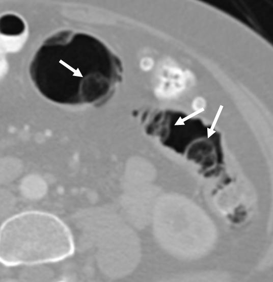
Fig. 6
Pulmonary and intestinal diseases are the most common benign causes of PI. Pneumatosis cystoides intestinalis (PCI) is a type of PI characterised by spherical gas collections in the bowel wall (arrows). PCI almost always occurs in the colon, and on colonoscopy it can mimic polyps. When viewed with CT colonoscopy, the gas cysts can be easily identified within the colon wall.
The most common life-threatening cause of pneumatosis intestinalis is bowel ischaemia. The combination of PI and an elevated serum lactic acid level is associated with a greater than 80% mortality rate.
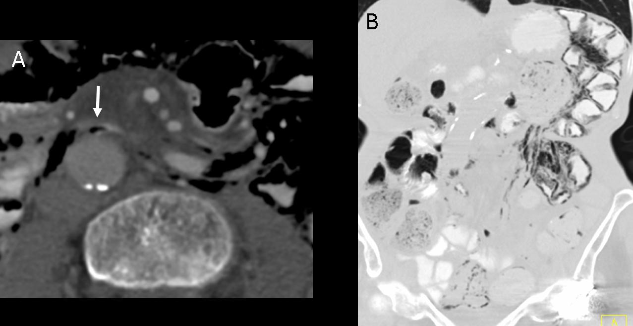
Fig. 7
CT shows gas within the mesenteric veins (white arrow in Fig. 7a). The distribution of PI is confined to the jejunum (Fig. 7b). When PI is confined to a portion of the small or large bowel within a specific vascular territory, ischaemia is the likely cause. Enteric capsule study demonstrated bowel ischaemia in this patient.
Pneumatosis secondary to infection
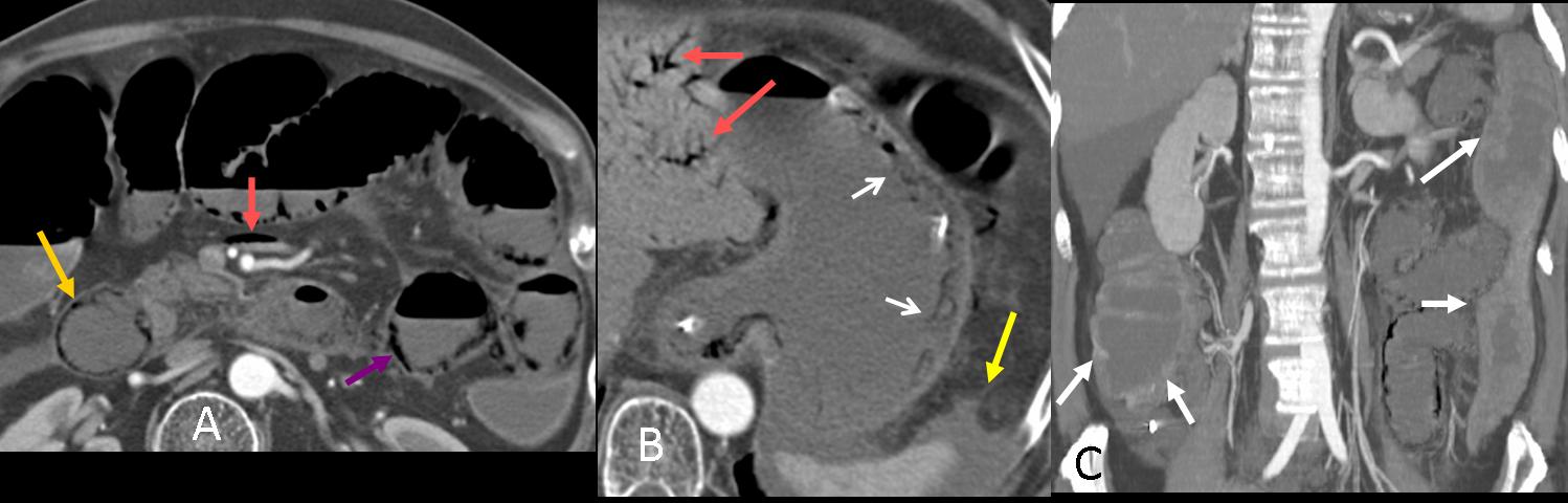
Fig. 8
PI in the duodenum (Fig. 8a, yellow arrow) and jejunum (8a, purple arrow) secondary to chemotherapy and infection in a patient with oesophageal neoplasm. In this case, gastric pneumatosis is also present (8b, white arrows). This PI distribution does not correspond to a specific vascular territory.
The presence of bowel wall thickening (white arrows), absent or intense mucosal enhancement, dilated bowel (8c, white arrows), arterial or venous occlusion, ascites (8b, yellow arrow), and hepatic portal or portomesenteric venous gas (8a and b, red arrows) increases the likelihood that pneumatosis has a life-threatening cause.
Portal venous gas
CT is more sensitive than radiography in detecting hepatic portal and portomesenteric venous gas, the presence of which increases the possibility of PI due to life-threatening causes. Portal venous gas (Fig. 9a, white arrows) can be differentiated from biliary gas (Fig. 9b, arrowheads) by its characteristic tubular branching lucencies that extend to the periphery of the liver, whereas biliary air is more central.

Fig. 9

Follow Dr. Pepe’s advice
Follow Dr. Pepe’s advice:
Pneumatosis intestinalis can be diagnosed in chest radiographs: look below the diaphragm when you read the film.
CT differentiates benign from life-threatening causes: cystic appearance suggests a benign condition. To diagnose life-threatening entities look for air in the mesenteric veins, clots within the mesenteric arteries, air in portal veins and additional bowel findings.
Suggested reading
- Pneumatosis Intestinalis in the Adult: Benign to Life-Threatening Causes. AJR 2007; 188:1604–1613.
- Pneumatosis intestinalis versus pseudo-pneumatosis: review of CT findings and differentiation. Insights Imaging. 2011;2:85-92.
Case prepared by Alberto Villanueva MD


bowel, chest, diagnosis, EDiR, European Diploma in Radiology