Since some of you misdiagnosed the right aortic arch in case 9, I will give you another chance with another mediastinal case in a 45-year-old man, who is asymptomatic.
1. Pericardial cyst
2. Pericardial fat pad
3. Thymic tumour
4. All of the above
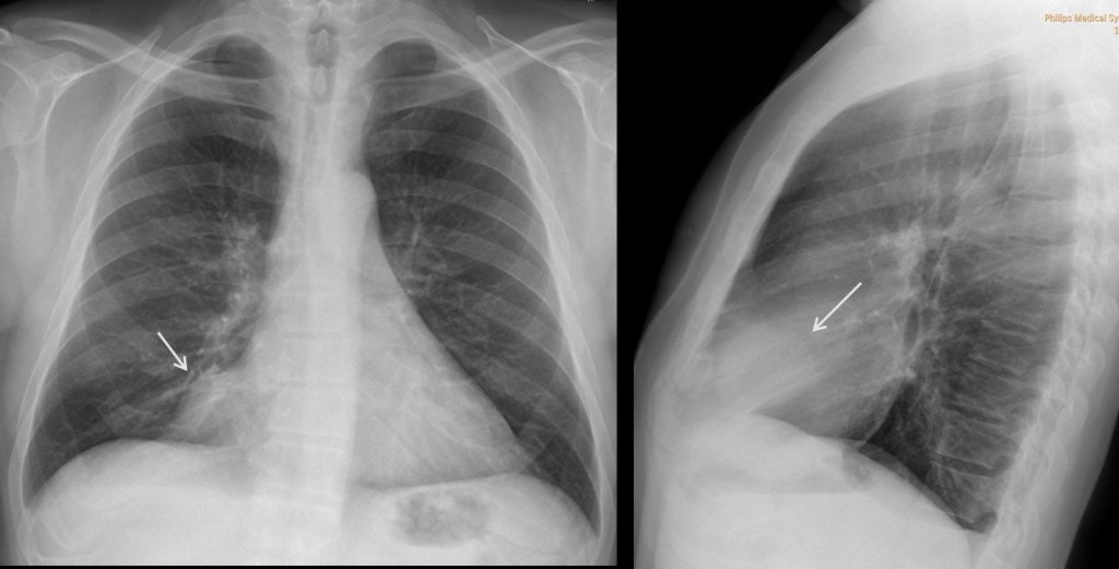
Fig. 2
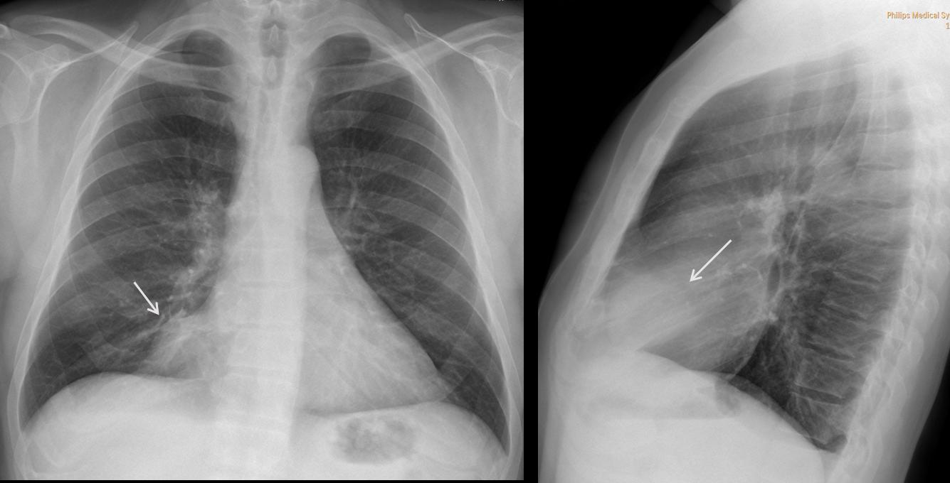
Findings: PA and lateral chest radiographs show an extrapulmonary mediastinal mass (arrows) occupying what is called the cardiophrenic space.
Masses in the cardiophrenic space (CS) are not uncommon and are easily recognized in the PA and lateral chest radiographs.
From the diagnostic viewpoint, all lesions in the CS have the same appearance on chest radiography and therefore, cannot be differentiated. The correct answer is: 4. All of the above.
On CT, these lesions can be of soft tissue density (mediastinal tumour), water density (pericardial cyst) or fat density (pericardial fat pad).
In the case shown, CT depicts a non-enhancing, rounded fluid-filled mass in the CS (arrow), measuring 7 H.U.
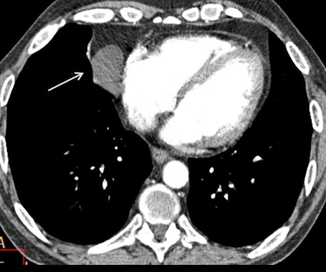
Fig. 3
Final diagnosis: pericardial cyst
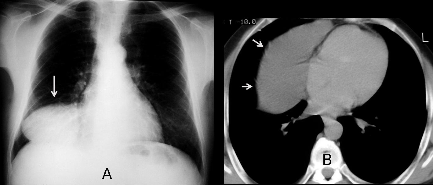
Pericardial cyst (Fig. 4, arrows) is the most common fluid-filled mass in the CS. It is located more commonly on the right than on the left and appears as a CS mass in the chest radiograph (Fig. 4a, arrow). CT shows a cystic lesion with well-defined borders and smooth walls (Fig. 4b, arrows). Note the change of shape in the upright and recumbent position, indicating a partially filled cyst.
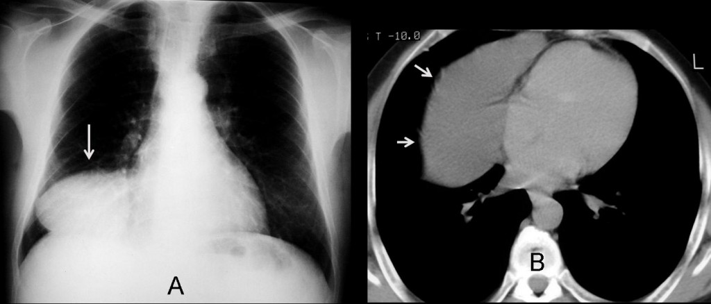
Fig. 4
Fatty lesions
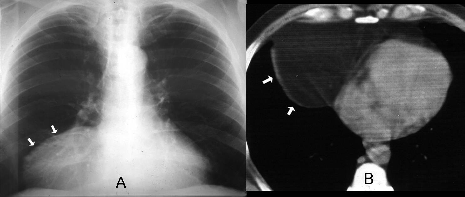
The most common fatty lesion in the CS is the pericardial fat pad. An enlarged fat pad can simulate a mass in the chest radiograph (Fig 5a, arrows). The diagnosis is easily made with CT, which confirms the large fat pad (Fig. 5b, arrows). The absence of encapsulation and lack of a soft-tissue component differentiate normal fat from the rare fat-containing tumour.
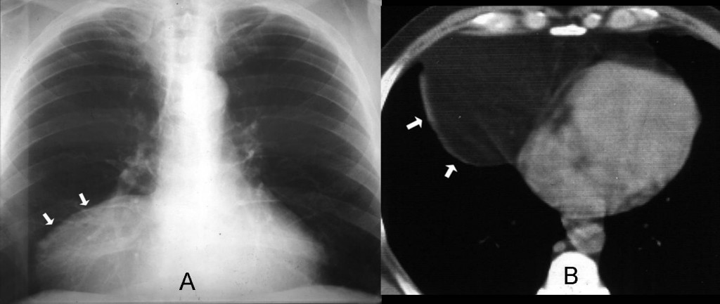
Fig. 5
Soft tissue density
The most common solid lesion in the CS is lymphadenopathy; they are usually multiple and not very large (Fig. 6, arrows). Lymphoma is the most frequent cause. They may also originate from tumors above or below the diaphragm.
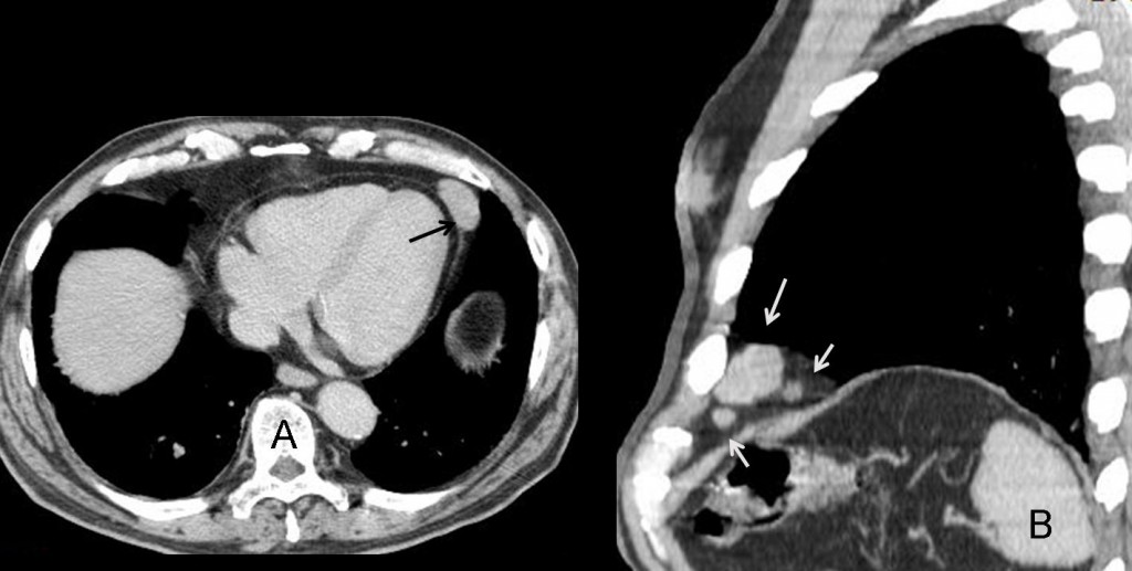
Fig. 6
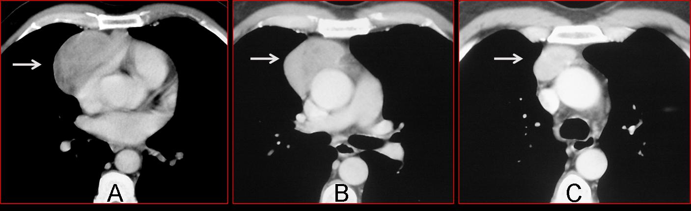
In large soft-tissue tumours of the CS, a thymic origin should be suspected. Although thymic tumours originate in the anterior superior mediastinum, they may slide down along the mediastinal planes and appear in the CS (Fig 7a+b, arrows). A useful diagnostic feature is that they always maintain a connection with the superior mediastinum, visible on CT (Fig 7c, arrow).
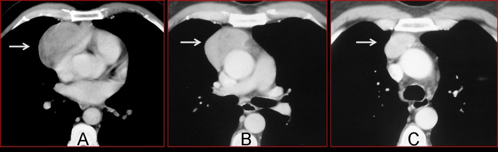
Fig. 7
Morgagni hernia
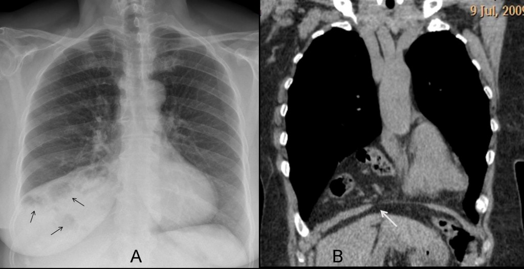
Fig. 8
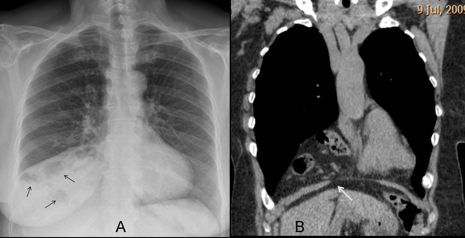
A hernia through the foramen of Morgagni may simulate a mass in the CS. The diagnosis can be suggested in the chest radiograph if aerated bowel loops are seen (Fig. 8a, arrows). If not, the diagnosis rests on CT, which shows the herniated fat and bowel and the diaphragmatic defect (Fig. 8b, arrow).

Follow Dr. Pepe’s advice
Follow Dr. Pepe’s advice:
Masses in the CS are not uncommon, and their etiology cannot be determined in the chest radiograph.
CT is diagnostic by showing the radiographic density: fat (pericardial fat pad, Morgagni’s hernia); water (pericardial cyst) and soft-tissue ( lymphadenopathy and the occasional thymic tumour).
Suggested reading: Lesions at the cardiophrenic space: findings at cross-sectional imaging. RadioGraphics 27:19-32. 2007
Case prepared by Dr. Pepe