
Dear Friends,
Here is case number 8:
Sixty-three year old male admitted to the Emergency Room with possible pulmonary embolism. Chest radiographs show a bulge in the descending aorta. Enhanced CT from the apex of the lungs to the diaphragmatic domes confirms the embolism. A more caudal axial CT shows a rounded opacity surrounding the aorta.
Diagnosis:
1. Neurofibromatosis
2. Lymphoma
3. Cystic lymphangioma
4. None of the above
Muppet is very frustrated about the previous case. He thought you would all diagnose it easily and says that if you don’t get the new one, he will retire to a nunnery…
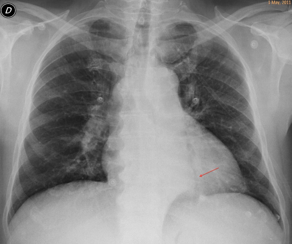
63 year old male, PA chest
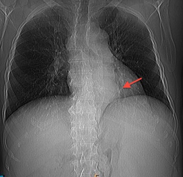
63 year old male, chest CT
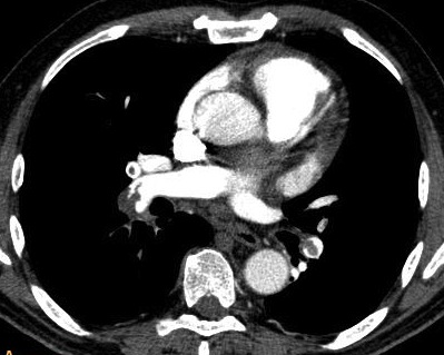
63 year old male, enhanced CT
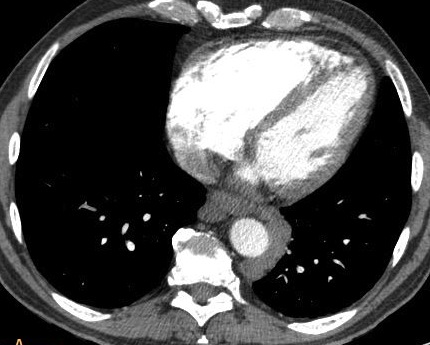
63 year old male, caudal CT
Click here for the answer to case #8
Aside from confirming the pulmonary embolism, CT reveals a rounded lesion around the aorta. The serpiginous appearance of the peri-aortic density suggests vascular structures. The prominent spleen and the enlarged azygos vein suggest portal hypertension with mediastinal varices. Diagnosis is confirmed with a delayed film, which shows enhancement of the varices (
arrows).
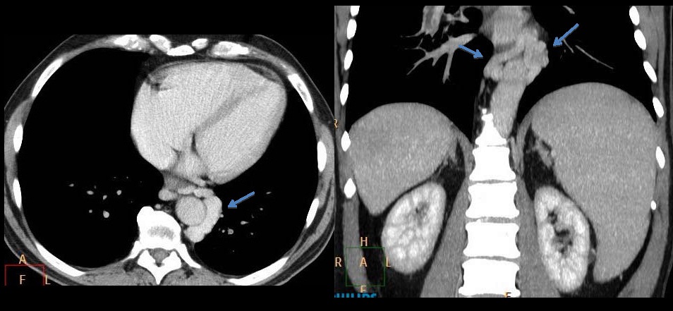
Answer Case 8
We found out later that this patient had chronic liver disease secondary to an extensive liver infarct years earlier. Mediastinal varices are not uncommon in chronic liver disease and are easily diagnosed by CT. In this particular case, because of the suspected pulmonary embolism, an early arterial phase CT was obtained, delaying the filling of the varicose veins.
Teaching point: the mediastinum is composed mainly of vascular structures. When a mediastinal abnormality is present, always rule out a vascular origin (arterial or venous).
Muppet wishes you the best for the coming year!


lymphoma
The axial CT-scan of the Thorax shows multiple infracarinal lymphnodes enlargement.
The second slice shows a paravertebral mass in the posterior mediastinum. The mass is of soft-tissue density and is surrounding the descending aorta without invasion of the visceral pleura (preserved fat plane). However, I suspect pressure effect on the vertebral body. There is no cystic degenerations or necrotic areas.
My DD would be:
1- Neurofibroamtosis.
2- Lymphoma
3- Extramedullary hematopoiesis
I would like to perform a CT-guided biopsy through parvetebral approach to confirm the diagnosis 🙂
Neurofibromatosis would be possible,but the mass shows no enhancement so extramedullary hematopoiesis
Lymphoma
Because of PE in presence of no other risk factors
Based on those images – Lymphoma. Because masses are confluent and PTE. Futher dx needs more images.
-Widening of left paraspinal line with lobulated retrocardiac opacity on chest radiograph with widened lower right paaratracheal stripe.
-CT chest showing pulmonary embolism with left paravetebral soft tissue with precarinal and retrocardiaac lymphadenopathy.
Lymphoma should be the first possibility.
Extramedullary hematopoiesis and mediastinal hematoma are other differentials but there is nothing in the history or on images to suggest these diagnoses.(presuming that thin linear structure seen in the descending thoracic aorta is artefactual,and not an intimal flap!)
there is volume loss involving left hemithorax. lateral view is necessary to assess lobar anatomy.
Why do I show the scout view of the chest?
Does it help?
It does help. Splenomegaly.
Consider the differential diagnosis of splenomegaly
did think about anomalous pulmonary drainage to the coronary sinus but images not convincing(probably ending up looking stupid saying this??). we need a good venous phase CT of chest and abd,
dilated azygous arch seecondary to caval obstruction ? hazardous to comment when there is inadequate opacification.
Extramedullary hematopoiesis would have know clinical history.. But the combination of enlarged lymph nodes i believe suggests lymphoma..
You’re cheating.. Scout is a (very very) devious way to show splenomegaly.. it is by far contextual as a finding on a scout, as there are other causes for lack of stomach air bubble. Given the fact you pushed splenomegaly, retrospectively that would cause trombocitopeny and PTE, so this plus splenomegaly would fit chronic myelogenous leukemia.
I have NEVER imagined i could diagnose CML on a chest X-ray. My humble respect, professor.
Thank you. My objective is to make you think and consider alternative diagnosis, other than the obvious ones. Yours is a good diagnosis; but still not the right one.
Wil give you a hint: why is the azygos vein enlarged in the PA chest film?
Bummer.. I’ll give it one more shot. Budd-Chiari syndrome? What really bothers me is that para-aortic mass does not really fit a lymph node station – there is none there (between pleura and descending aorta); i’ve first said lymphoma because of encasing character. Are those some collaterals in azygos/ hemiazygos system?
Merry Christmas !!!
The air bubble is present and displaced towards the right. That, and the opacity in the left upper quadrant suggests splenomegaly.
I am not cheating. Muppet is 😉
I vote for four! Greetings for Mr. Muppet.
You are right. Muppet congratulates you 😉
But, still, nobody has mentioned the right diagnosis.
The soft tissue surrounding the descending Aorta is an abnormal juction probably between the Hemiazygos and azygos vein, witch are clearly enlarged.
These foundings could suggest an obstructive proces of the inferior vena cava and probably a neoplastic proces in the abdomen- liver?
thoracic splenule
? left side IVC with hemiazygos continuation
can be loculated pericardial/pleural fluid
? venous anomaly of hemiazygos system
Left sided IVC with hemiazygos continuation
Deep veins thrombosis,Neo pancreatis?paraneo sy.?
IVC thrombosis and Cavo-caval anastomosis-shunt true azygos-hemiazygos system…other diagnosis consider esophageal varices in cirhosis,but there is no signs of superficial colaterals-caput medusae?splenomegalia is present…maybe meta in hepate,or other some short-term pathology?
No hereditary vascular malformation,no evidence of colaterals
OK, after the azygos tip, you all are close enough. Will give you a collective A! ( Muppet wanted to scale it down, but I stayed firm). Wait for the final answer.
now it is clear that there is a process of venous shunt through azygos system , but what about the translucent line retrocardiac , is it the esofagus displaced laterally by something which also obstruct the IVC ? the fact o splenomegaly and these shunts let us think about a problem that needed time for all these modifications to appear , and the association of pulmonary embolism again let us think about systemic cause of general dissiminated thrombosis , i agree with the idea of paraneoplastic sdr. regarding the age of the patient , thrombosis may be also involve the supra hepatic veins and this is the Budd Chiarri sdr. ?? .
Lymphoma
Retroperitoneal fibrosis causing portal vein hypertension – therefore as a consequence – a dilated azygos and splenomegaly.
This was the best case of all!!! Happy 2012!
wonderfull case
Oesophageal varices ..dilated azygous vein secondary to portal hypertention ..with retrocardiac mass …represent oesophageal varices.