Dear Friends,
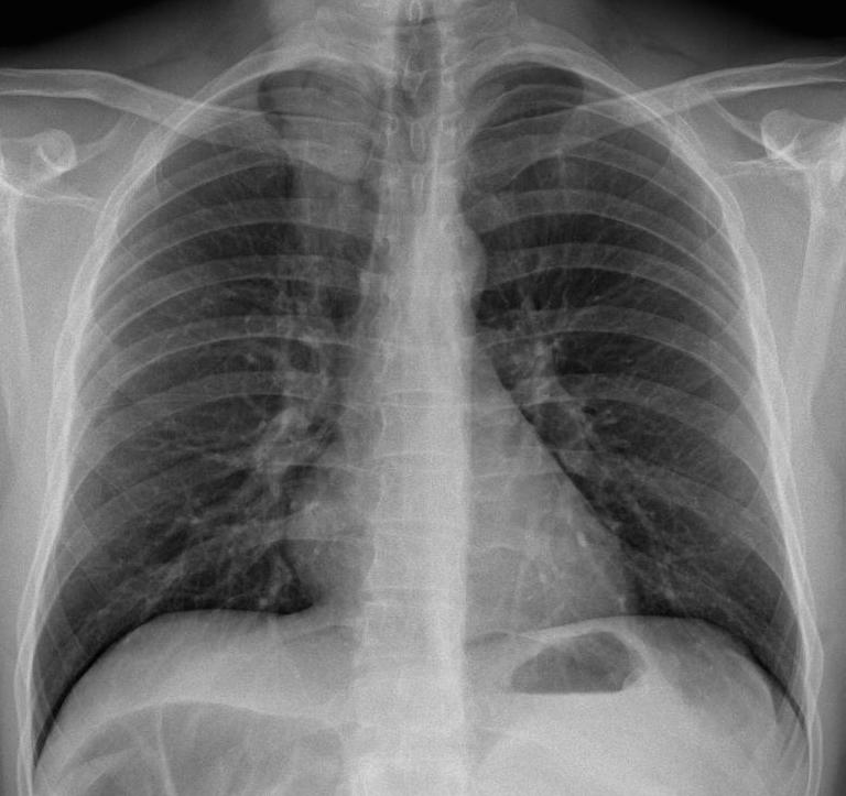
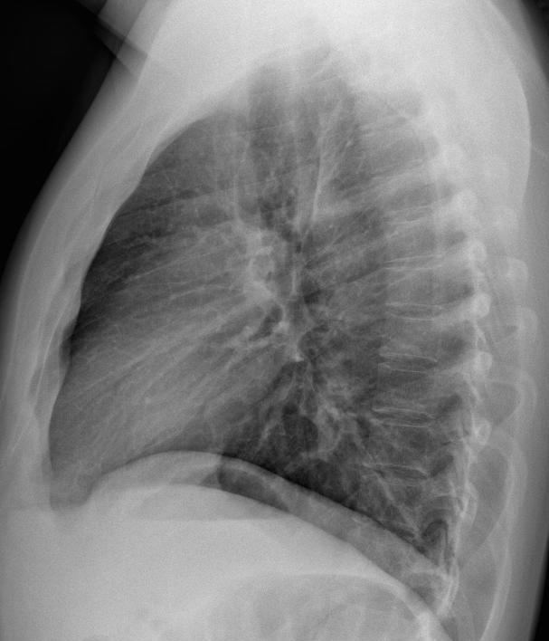
Muppet insists on showing the following case that he saw recently: preoperative chest radiographs of a 25-year-old male with seminoma. Check the images below, leave us your thoughts in the comments, and come back on Friday for the answer.
Diagnosis:
1. Tuberculosis
2. Metastases
3. Mucous impaction
4. None of the above
Click here for the answer to case #109
Findings: PA chest radiograph shows a triangular opacity in the right upper lung (A, arrows). The appearance is unspecific. The clue to the diagnosis lies in discovering that the opacity extends into the soft tissues of the neck (A, red arrow), indicating that it is not located in the lung and originates outside the chest.
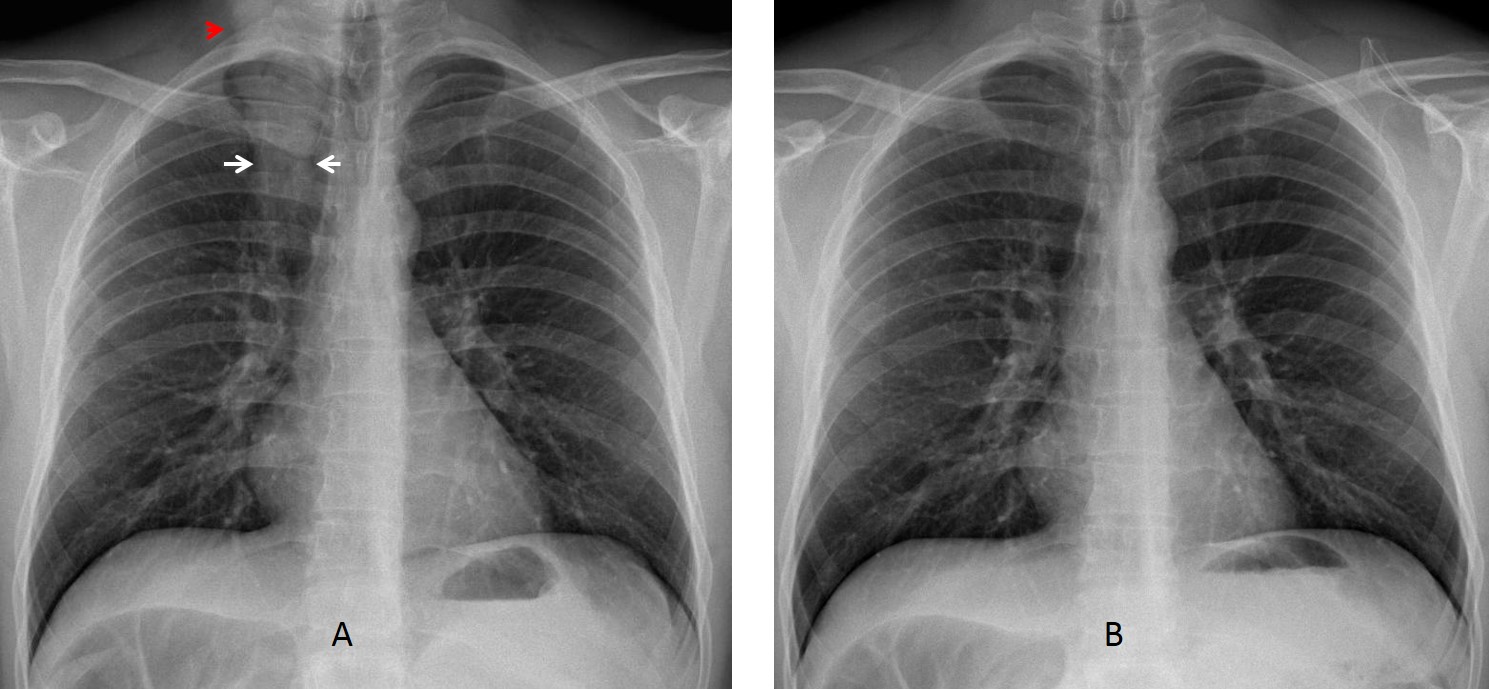
Examination of the patient showed a long ponytail. A new radiograph after lifting the ponytail (B) confirms it as the cause of the opacity.
Final diagnosis: ponytail simulating lung disease.
Congratulations to David who was the first to answer and to give the correct diagnosis.
Teaching point: always remember that long hair may simulate apical lung disease.


I think that is something extrathoracic, like tail hair. In lateral view is not visible and probably is localized at skin surface of the posterior region of the thorax.
The answer 4: none of the above.
I agree, in PA view you can see it reaches above thoracic inlet.
I agree,looks like braided hair artefact…
Pleural based mets?
2. Metastases
Neck mass extending posterior to the spine. ? Metastases from the seminoma or extragonadal seminoma.
Opacity with ill defined borders at the upper part of the right lung in the PA view.
In the lateral view it seems to be extrathoracic, with well defined borders posteriorly.
Because of its size and location, I would suggest elastofibroma of the back.
Elastofibroma is a chest wall lesion and it is not visible in the plain film. Sorry.
I am sorry, too! It looks like braided hair in the PA view, but why so well defined borders in the lateral?
To me, the shadow in the lateral view looks like the scapula. But I am not infalible (working on it, though).
Agree with David. Seems like mine when I was young.
You are still young
definitely extrathorasic -hair tail!
Is it subcutaneous lipoma of the paravertebral area?
This density is definitely extra thoracic, extending to the soft tissue of the neck.
Alongside the density borders and some focal spots inside the density me can see encaged hyperlucent areas (Air?)
i think is somethink like dermal( mole? ) or extra dermal artifact lesion ( hair? )
plus Chilaiditi syndrome
metastasis pleural de osteosarcoma
The opacity in the upper part of the right lung on the PA view is seen it extends upwards to the neck with well defined margins except the lower parts and the right margin cranially, opacity is inhomogenous with hiperlucent zones, I do not see the opacity on the lateral view. I suggest it is extrathoracic and artificial.
Chilaiditi syndrome.
…la prima cosa che farei è’ richiamare il paziente e farei un esame “ispettivo”…..ricordandomi che sono innanzitutto un…..clinico……all’esame ispettivo, farei seguito quello ‘palpatorio’….. Aiutandomi con la palpazione ” digitale” , vale a dire una ecografia………risposta finale 4. ….potrebbe essere una patologia dei tessuti molli del collo o il risultato di una FNAB….con stima da Bari …..
4. None of the above
4. None of the above