Today I am presenting chest radiographs of a 60-year-old man with vague chest complaints and mild cough.
What do you see in the images below? Leave your thoughts in the comments section and come back on Friday for the answer.
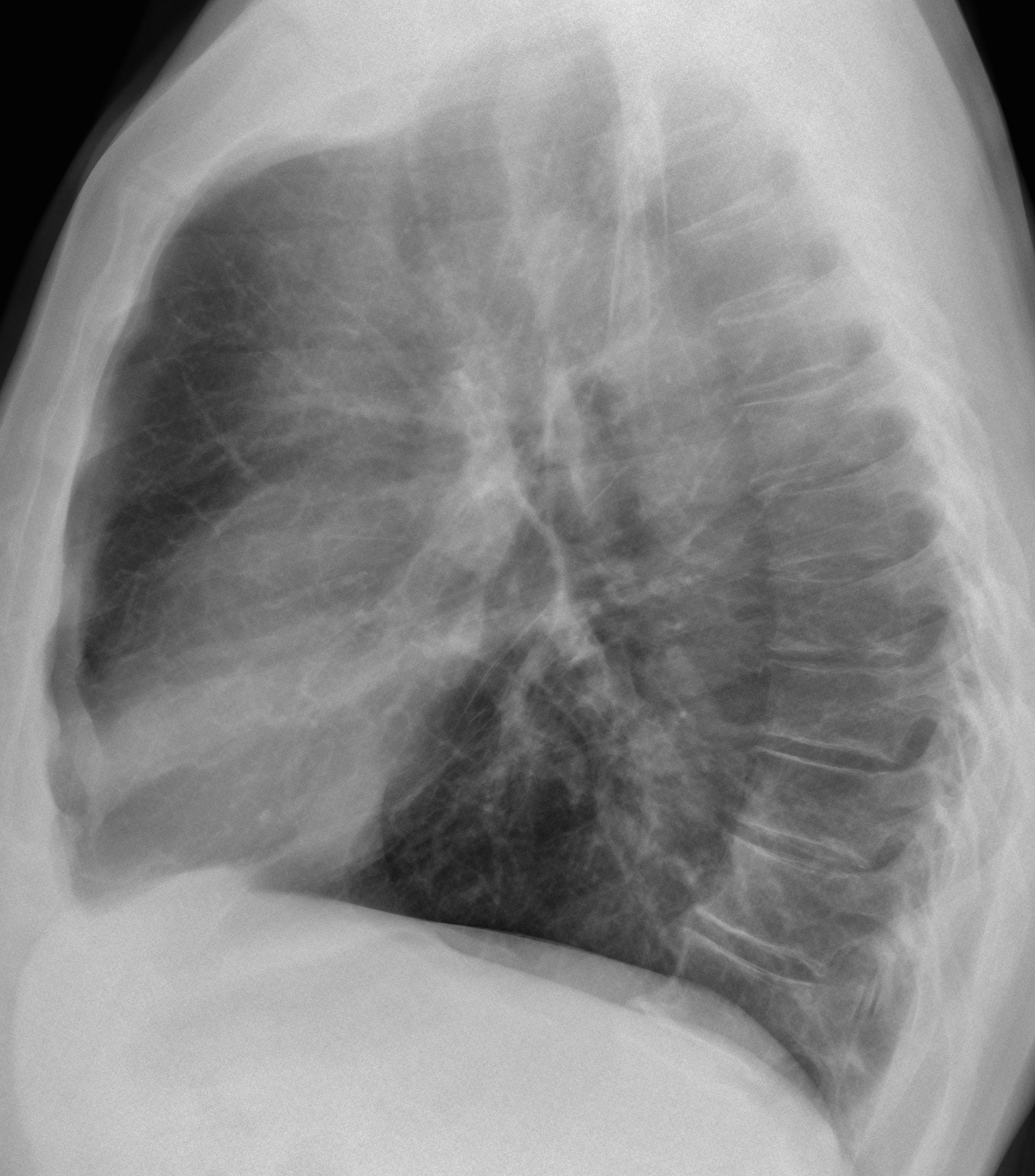
Findings: PA radiograph shows a lung nodule projected over the anterior left first rib, also visible in the lateral view (Fig 1, arrows).
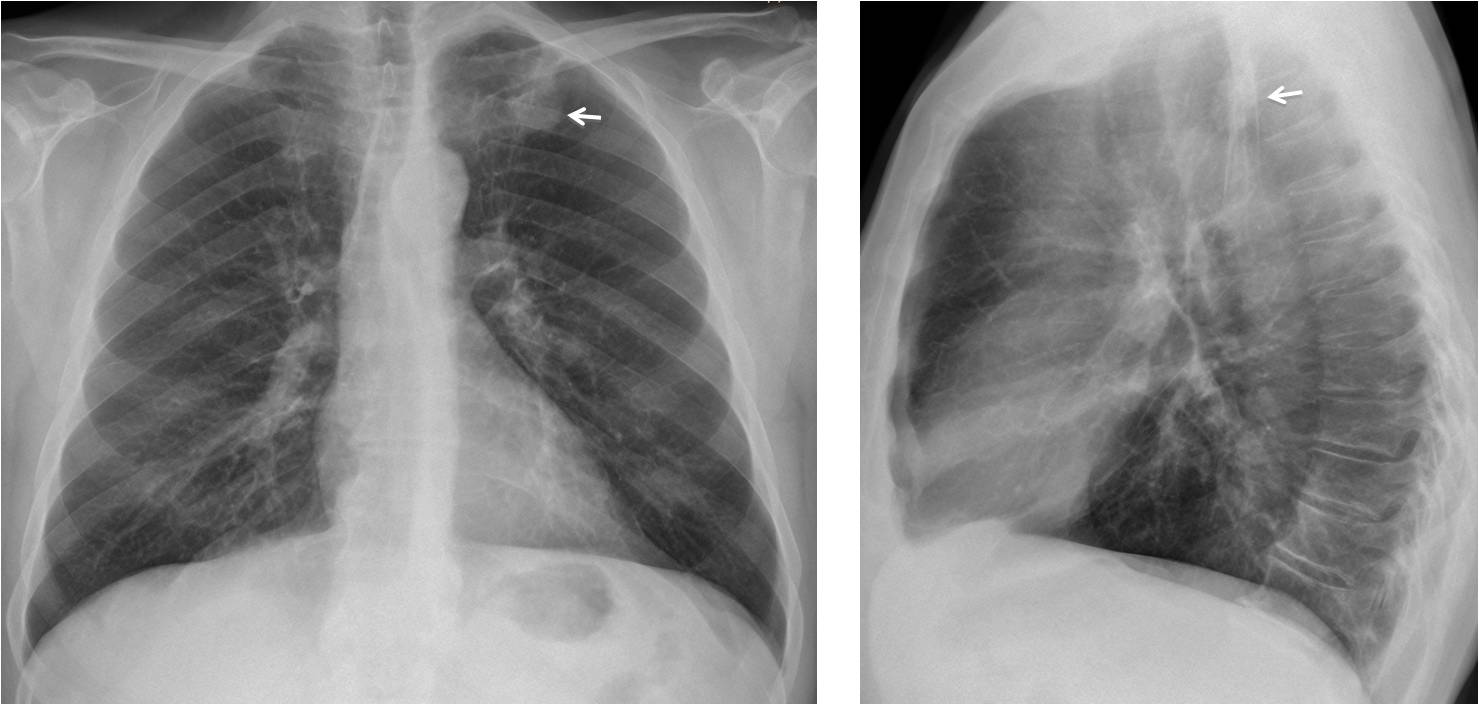
Fig. 1
The nodule was barely visible in films taken four months earlier (Fig 2, arrows).

Fig. 2
The nodule was missed in both studies. Radiographs taken 18 months later show a large mass in the LUL and a right perihilar mass (Fig 3). Bronchoscopy and biopsy confirmed carcinoma in both locations.
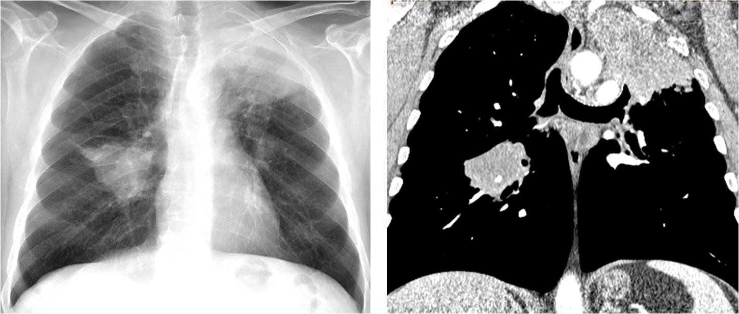
Fig. 3
Final diagnosis: fast-growing bilateral lung carcinoma
Congratulations to all of you who discovered the nodule, with special mention to Genchi Bari, who was the first to mention it.
I am showing this case to emphasise the importance of scrutinising the lung apices. The reported miss rate of nodules in this location reaches up to 50% and many of them are carcinomas.
Teaching point: always look at the apices. This patient is living proof of what happens when a nodule is overlooked.


….tumore di Pancoast , a sx, sino a prova contraria.
downward displacement of the right hilum but no clearly evidence of atelectasis.
tubular opacity on the right pul.base (vascular etiology?)
i think i see a mass and not artifact on the left First sternum rib join
maybe upward displacement of the left hilum 🙂
opacity at the apex of the left lung
Left apical very dense nodule likely calcified and pleural thickening. TBC related? It seems like if there were some lucencies protected over this apical nodule.
Not certain but possible lung nodules in left lower
The upper porto on of tracheoesophageal stripe is thickened. Any oesophageal disease?
No esophageal disease
1. Dense nodule i the left lung apex, best seen on lateral image.
2. Possible, small nodule in the proximity of the left hilum.
3. Tubular lina in the right costophrenic angle- might intrathoracic rib or aberrant vessel or unusal presentation of a regular vessel.
Do We deserve another hint?
No hints. You already mentioned the abnormality
1. Intrapulmonary mass in the left lung apex.
2. A small left parahilar nodule, lateral to the descending pulmonary artery.
A rounded mass lesion on left apex of lung. Right hilum seems to be displaced downwards (or is the left hilum displaced upwards?). Is there a mass just below the left hilum displacing it up? Im not sure.
Please enlighten.
The hila have not changed in comparison with a previous film. If I had to bet, my opinion would be that the left hilum is slightly elevated (but unchanged).
Sending you the previous PA an lateral.
Se observa un nódulo pulmonar en el ápice izquierdo, además se aprecia un desplazamiento anterior de la traquea , visto en la proyección lateral, con un aumento de la densidad a ese nivel. El hilio izquierdo esta desplazado hacia arriba ligeramente. Hay una destrucción osea del arco posterior de la tercera cotilla izquierda. La impresión es que es un cáncer de pulmón con mets osea y adenopatía paratraqueal.
Aunque no se ve muy bien en la placa PA, la tercera costilla está intacta. El nódulo es real.
nodule apical gauche; je demanderais un scanner thoracique
You are right. Unfortunately, the nodule was not seen at that time.
aspergiloma
..carissimo professore , grazie per la menzione….mi solleva il morale , dal momento che il mio Bari è stato travolto a Crotone , per 4 ad 1…..come e con lo stesso risultato del Barca !!!!!!.
Yes, we are brothers in sorrow…
nodule left apex