Today we are showing radiographs of a 59-year-old man with chest pain. What do you see?
Check the images below, leave your thoughts in the comments section, and come back on Friday for the answer.
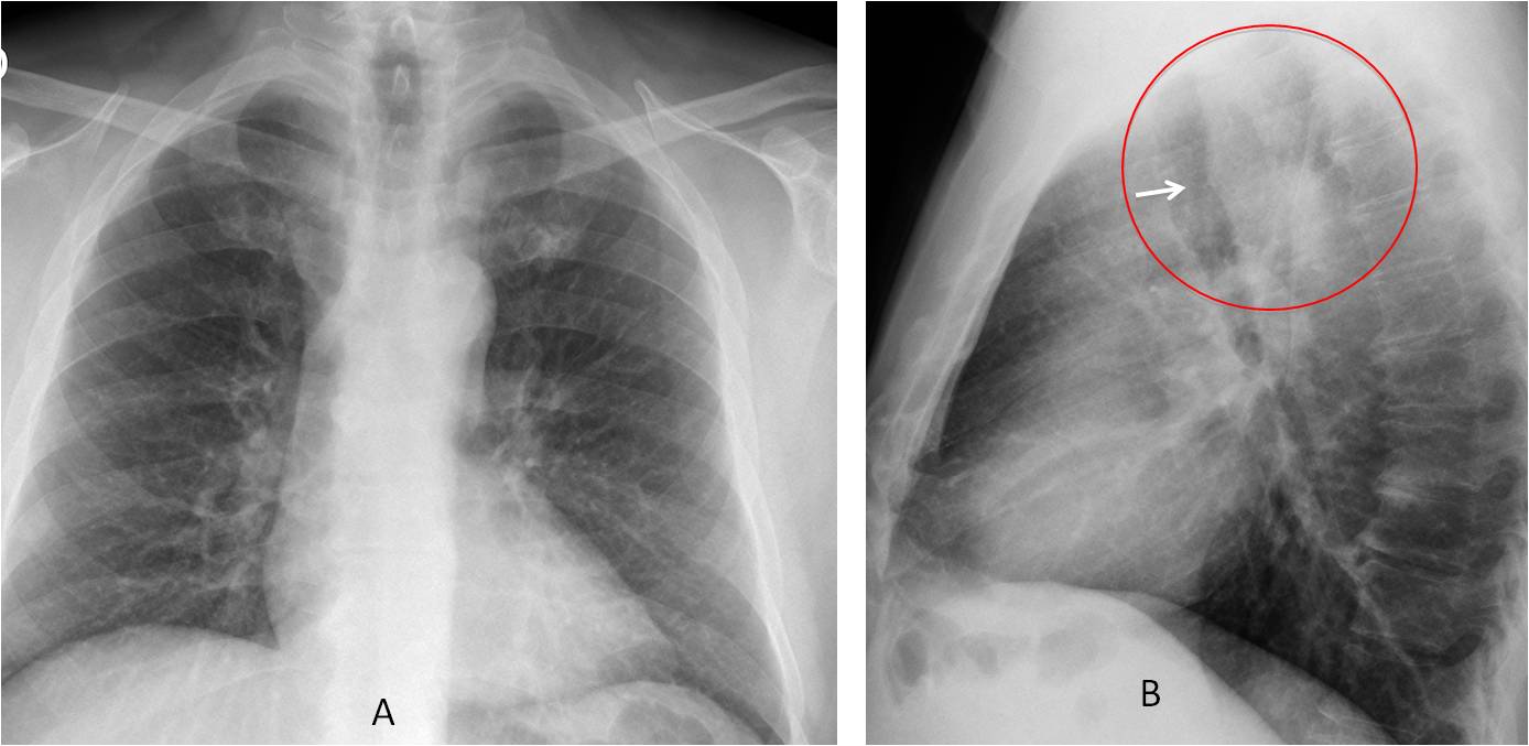
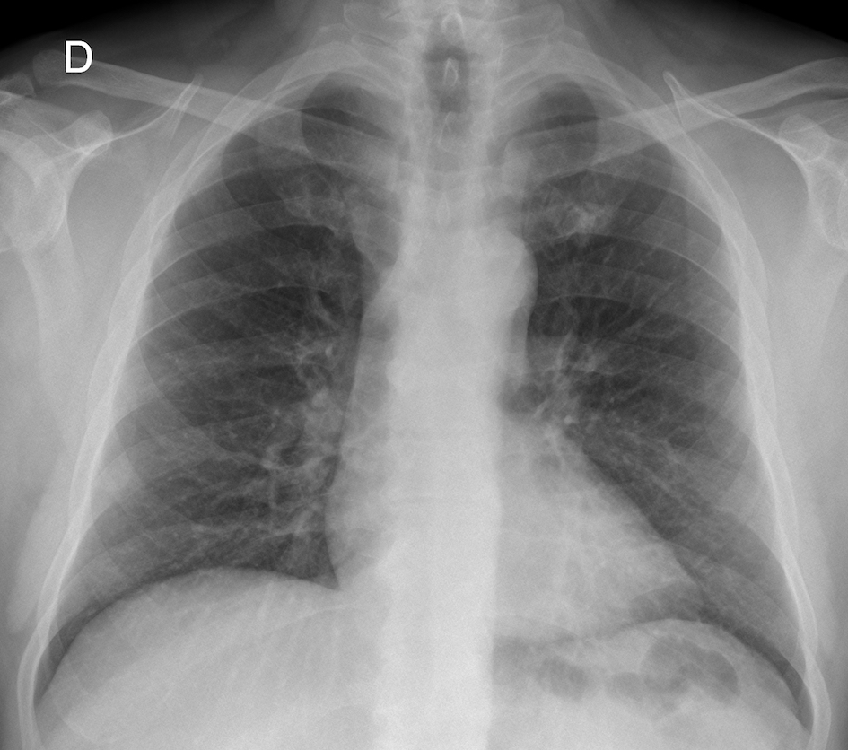
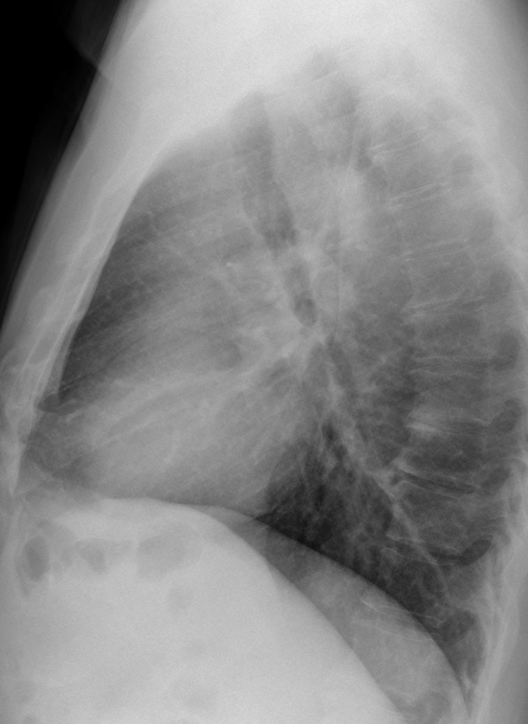
Findings: the PA radiograph (A) is unremarkable. The lateral view shows a slight protrusion of the posterior tracheal wall (B, arrow), accompanied by increased opacity of the retrotracheal space (B, circle) with disappearance of the posterior paratracheal line. These findings are highly suspicious of a posterior mass (see
Dr Pepe’s Diploma case no. 72), most likely arising from the oesophagus. Vascular malformation is less likely in the presence of pain.
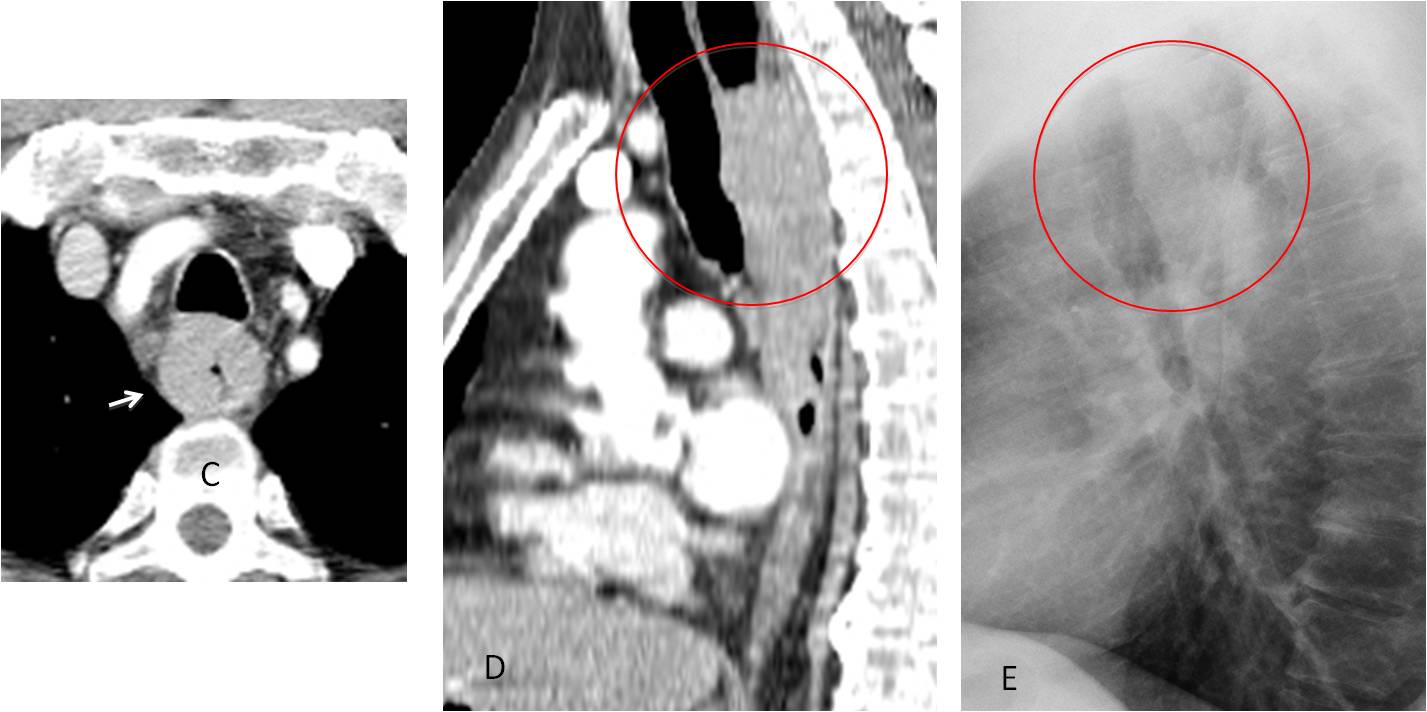
Coronal CT shows a retrotracheal mass (C, arrow). Sagittal CT reconstruction mimics the findings in the lateral view (D-E, circle).
Final diagnosis: carcinoma of oesophagus
I showed this case to emphasise the importance of the trachea and retrotracheal space in the lateral view. All of you who mentioned abnormalities in this area will get a Gold Medal, with special mention to Ivan.
Teaching point: always remember to look at the trachea in the lateral view. It may offer important information, as in the present case.


dilatation of pulmonary vessels
Pleural tickening in the upper lobes, more evident at right.
double contour of the aortic knob and the descending aorta. Suspicious for aortic dissection, given the chest pain of the patient.
I like this idea…
I think same
On lateral X ray i can see some blastic lesions on the vertebrae. Maybe malignancy related?
The sclerotic findings are secondary to degenerative changes. CT did not show any blastic metastases
Aberrant right subclavian artery
What do you see?
anterior tracheal displacement
suspicious retrocardiac mass
What are the causes of anterior tracheal displacement?
esophageal mass
Aberrant right subclavian artery between oesophagus and trachea
Aberrant subclavian goes behind the esophagus. I believe you are thinking of aberrant left pulmonary artery (pulmonary sling)
morgagni hernia?
Something more serious
…vi è una impronta, vascolare, dietro la trachea in LL…quindi una anomalia vascolare di decorso dell’aorta( destroposta ?) o dei suoi rami collaterali….di più non si può dire , dai radiogrammi standard…
I see hyperlucency above left diaphragm: maybe rapture of diaphragm?
Very unlikely. No previous trauma.
Posterior superior mediastinal mass? Does he have dysphagia?
Yes, he does.
soft opacity between trachea and esophagus, pushing trachea forward, indenting posterior margin.
can aberrant left pulm artery be that large?
Round opacity right paratracheal region?
Aberrant left pulmonary artery is lower
tumor mass located in the middle mediastinum ( superior).
+ is suspected the zone of calcification of cartilage of the first rib on the left.
lateral view:
posterior indintation of tracheal wall due to posterior course of aberrant left subclavian artery