Considering that this is Holy Week, I want you to do penance. This week’s images belong to a 73-year-old woman with mild dyspnoea and vague chest complaints.
What do you see? Check the images below, leave your thoughts in the comments section and come back on Friday for the answer.
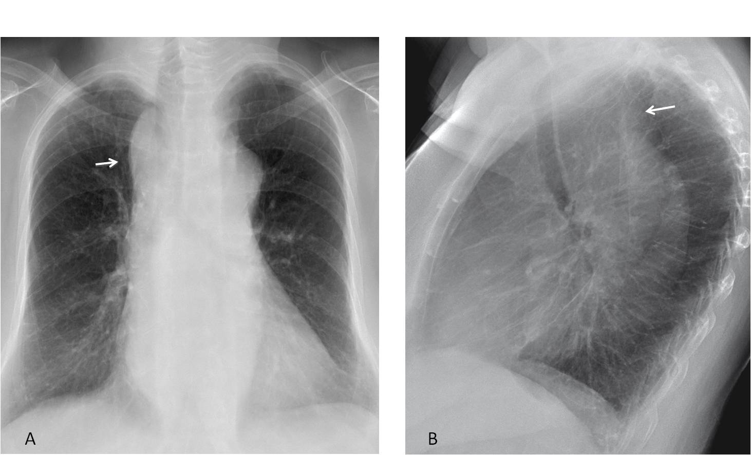
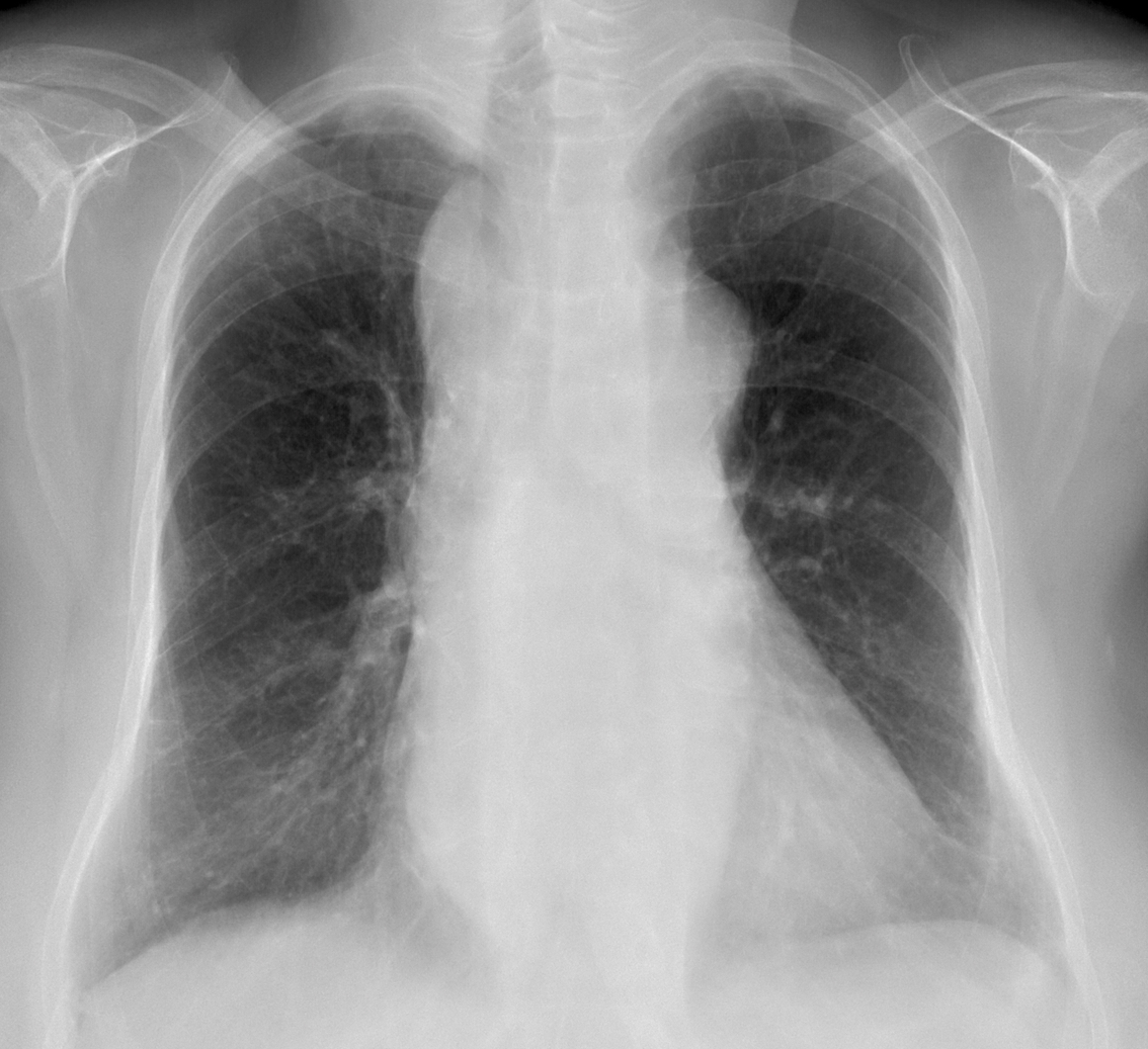
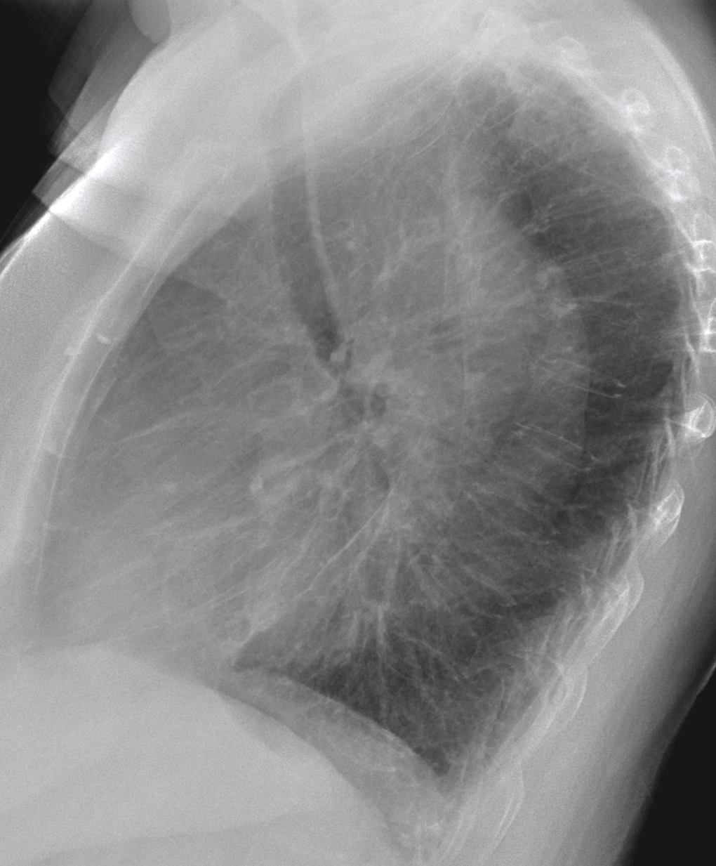
Findings: the PA radiograph shows widening of the right superior mediastinum (A, arrow), which in the lateral view is located behind the trachea (B, arrow). The initial impression is of an upper middle mediastinal mass. The first diagnoses that come to mind are a goiter or a dilated aorta.
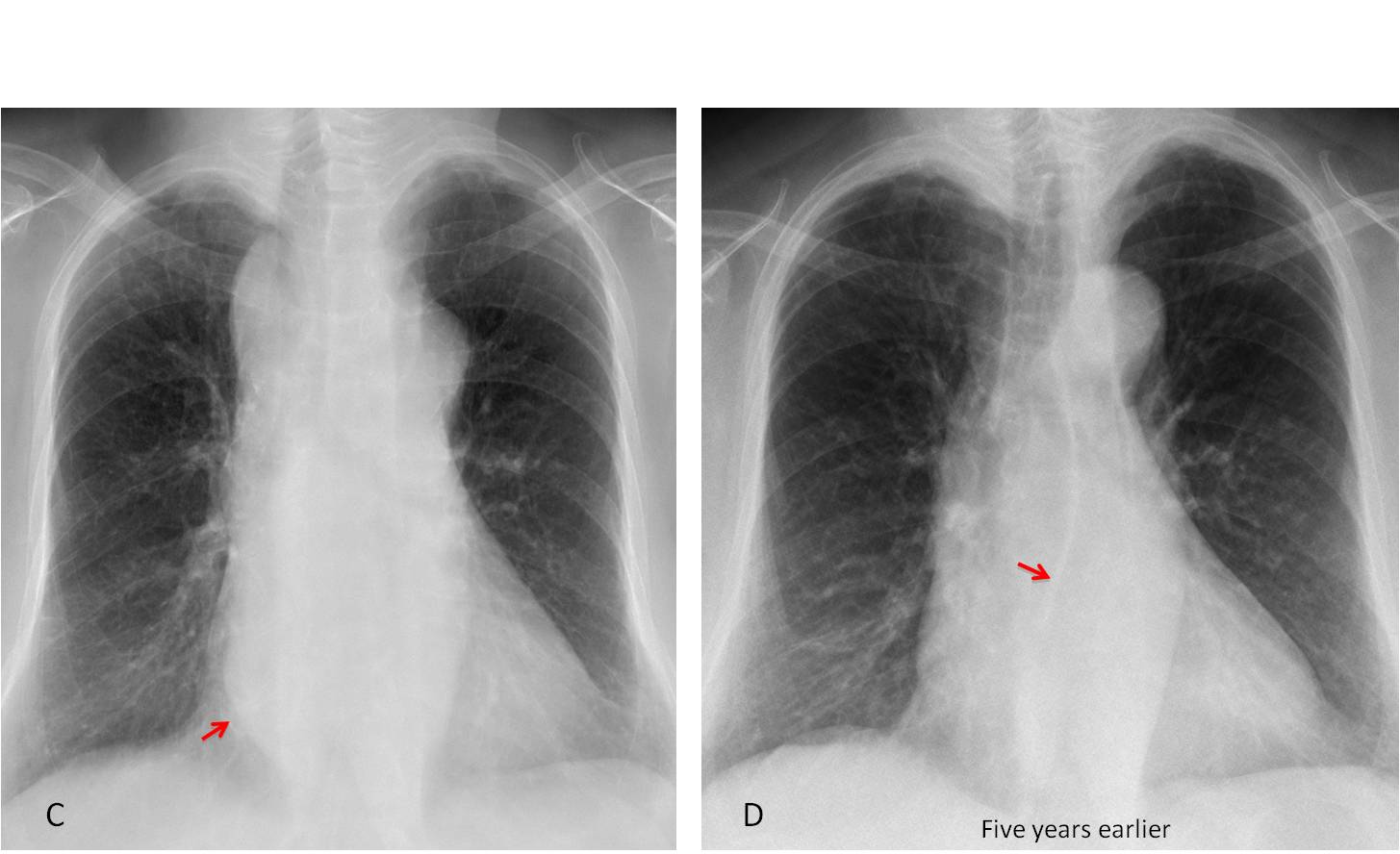
However, this is a typical case of satisfaction of search (again!). Most of you have missed the bulging of the azygo-oesophageal line (C, arrow), which is more evident compared with a film taken five years earlier (D, arrow). Therefore, we are dealing with a mass that extends along the middle mediastinum from top to bottom. The findings point to a dilated oesophagus or, more rarely, to a dilated aorta.
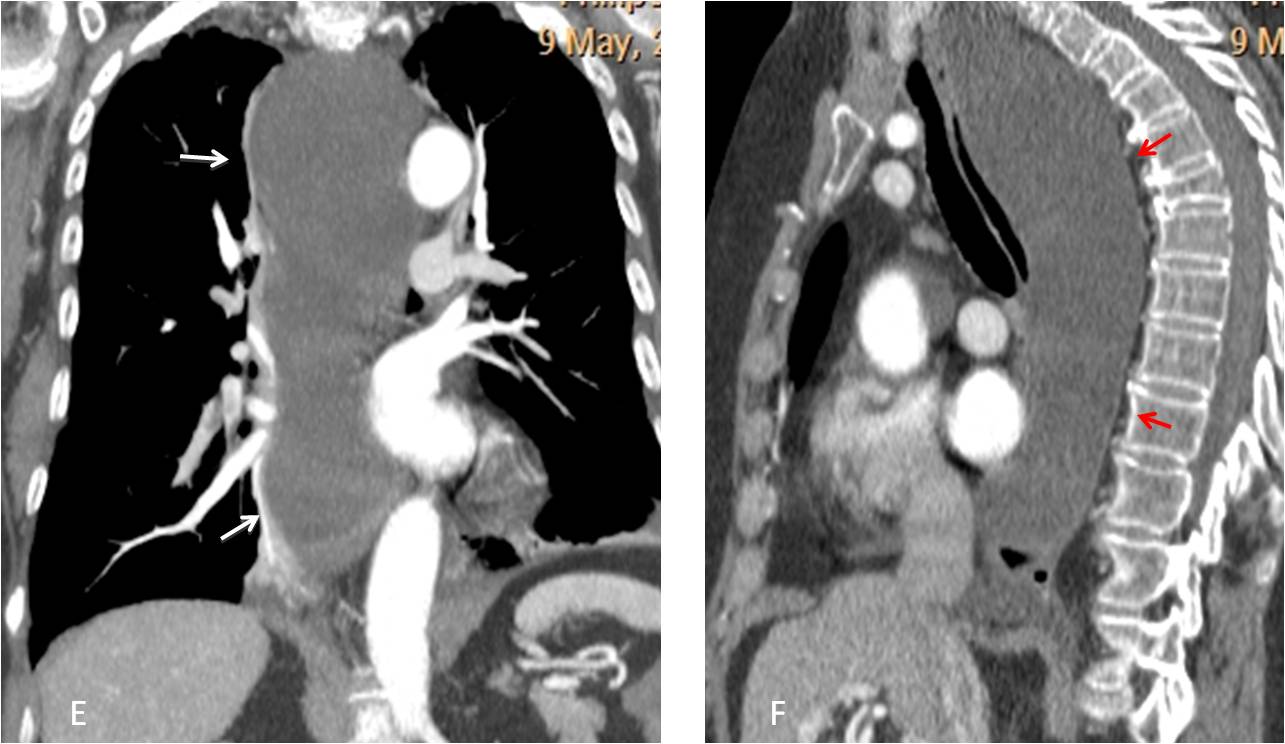
Oesophagogram was unremarkable. Coronal and sagittal CT shows a cystic tubular mass extending along the posterior wall of the oesophagus (E-F, arrows).
Final diagnosis: cystic lymphangioma of mediastinum.
This is a difficult case and I didn’t expect you to make the diagnosis. But I believe that you should have noticed the bulging of the azygo-oesophageal line and suggested a dilated oesophagus, which is the most sensible diagnosis.
Kudos goes to Bujar, who was the one to get closer to the truth.
Teaching point: Failing to look beyond the obvious is the cause of about 20% of misdiagnosis.


There is increased width of mediastinum shadow well defined on frontal and lateral view, suggesting additional mass.
Probable tymoma
On lateral view – there are:
-trachea ventral disposition – it seems like this mass is also in posterior part of mediastinum
-regarding the old age it could be also goiter – because of mass effect on airways
-compression fracture of the thoracic vertebrae, probable osteoporosis cause
So, there is additional mass in mediastinum, with positive mass effect on airways, old age
It could be thymoma, but more it looks like goiter
so-called posterior descending goiters
Intrathoracic goiter, compression fracture of the thoracic vertebrae
Hello,
there is oppacification of anterior retrosternal space which correspond to well delineated mass in upper mediastinum on ap view.
I consider 4T in DDx of anterior mediastinum or vascualar origin of change.
Hola Dr.
En mi valoración de la radiografía veo un ensanchamiento del mediastino, con desplazamiento ventral de la traquea. Me impresiona sea de causa vascular. ¿Tendrá un aneurisma de la aorta ascendente?
Saludos
Yunia (la cubana, residente de familia, 🙂 )
…pregiatissimo professore , a me, la massa sembra nel mediastino medio, per la compressione e dislocazione in avanti della colonna d’aria tracheale e la dislocazione posteriore dell’esofago…pertanto la massa potrebbe essere di natura adenopatica ( Castleman?).. .ho sbagliato tante volte quest’anno e probabilmente anche questa volta: la mia ” penitenza” la farò a SIVIGLIA, partecipando alle processioni della settimana santa…..Buona Pasqua ed un abbraccio dall'” ANDALUSIA “!
A believe la Semana Santa en Sevilla is an unique event. Enjoy your time in my city!
Widened mediastinum with increased retrocardial opacity funneling towards diaphragm. Lateral shows opacity in anterior mediastinum.Tracheal column is shifted to the right and anteriorly. There is pleural effusion of posterior phrenicocostal sinus on the right., better seen on profile.Aneurysm of ascending aorta, aortic arch and descending aorta would be my first guess. There is also reduced vertical diameter of ThV with hyperkyphosis and reduced mineralization of bones.
Do you have any alternative diagnosis?
Yes. Hiatal hernia.
Entire mediastinal widening with double cardiac density. Excluding this pacient anamnestic simptoms, my opinion as diff.dg is esophageal achalasia.
Widened mediastinum with anterior displacement of trachea so mass is posterior mediastinal with double density of the aortic arch on lateral view so mass may encroach on middle mediastinum. DD broncogenic cyst
Esophagenal tumor
Aortic aneurysm
Εsophageal achalasia
Double aortic arch
double aortic arch with vascular ring