Dear Friends,
April is the cruelest month. Dr. Pepe has eloped with Miss Piggy and has not prepared any Diploma cases. To cover for him I have selected five cases to be shown during April. Paraphrasing the title of a film by Jack Nicholson, I am calling them “Five easy pieces”. All of them show subtle findings, easily seen if you look carefully.
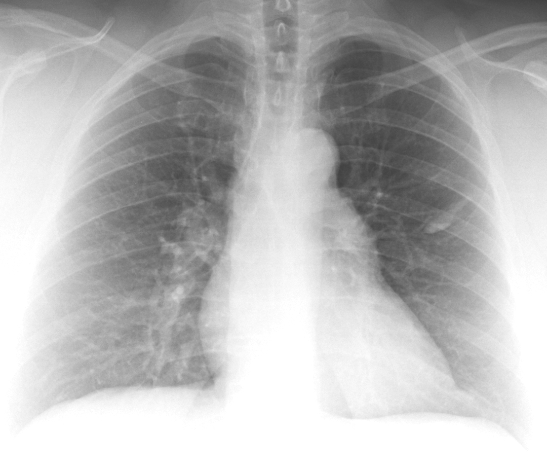
The first case is a PA radiograph of a 53-year-old woman, preo-op for bariatric surgery. Check the image below, leave me your thoughts in the comments section, and come back on Friday for the answer.
Click here for the answer to case #134
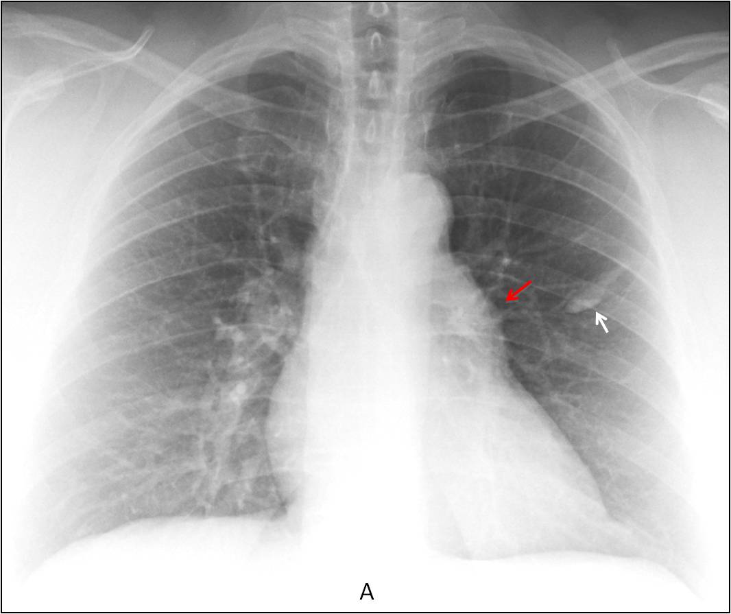
Findings: PA radiograph shows a partially calcified elongated image in the left middle lung field (A, arrow). Milk of calcium, as suggested by Borsuk, cannot be excluded. There is convexity of the pulmonary arch (A, red arrow) that was overlooked in the initial reading. This finding is compatible with enlargement of the main pulmonary artery or a mediastinal mass (See
Dr. Pepe’s Diploma Casebook case #52).
Unenhanced sagittal and axial CT shows a partially calcified mucous impaction (B-C, arrows) and an unexpected solid anterior mediastinal mass (C, red arrow).
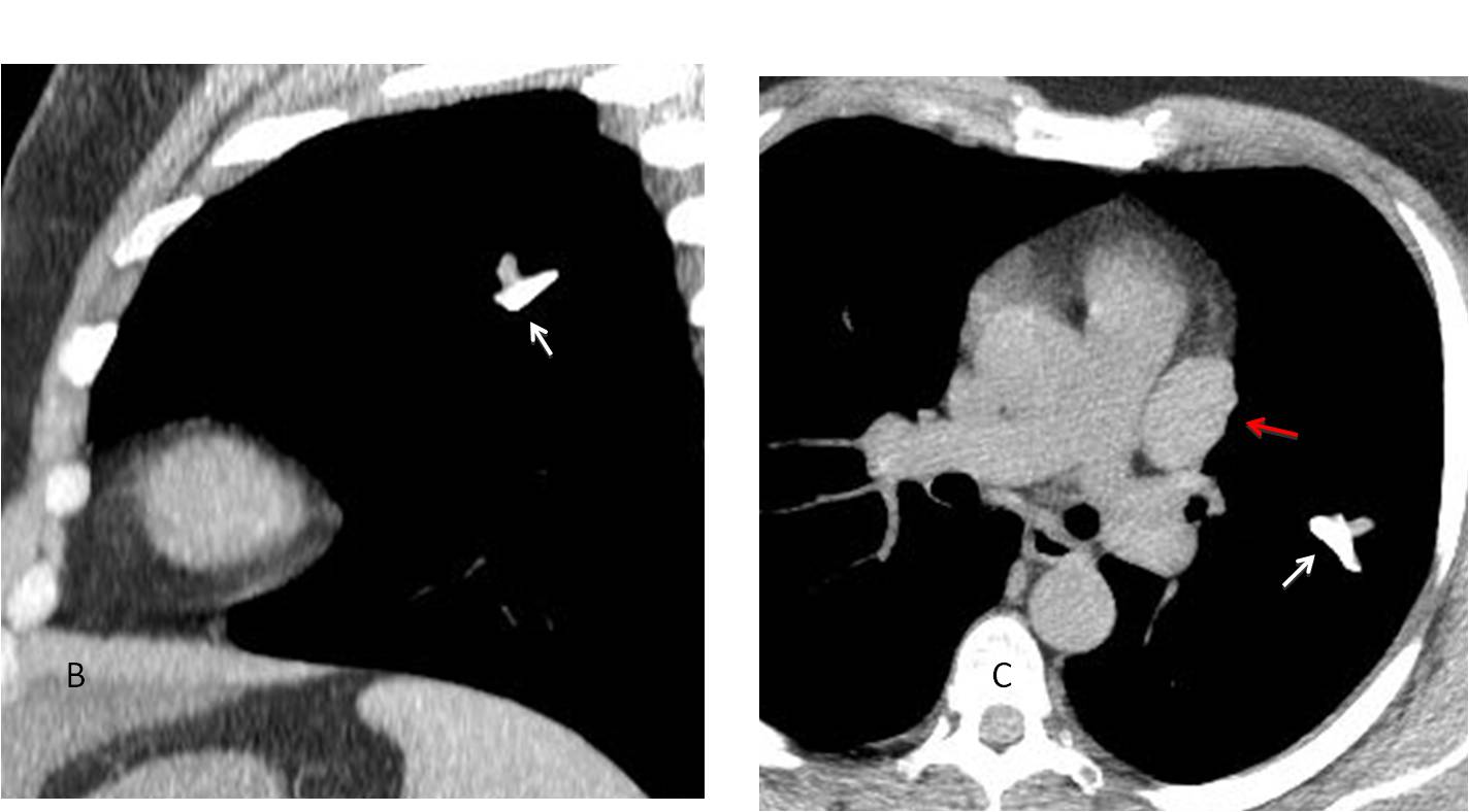
Final diagnosis: calcified mucous impaction and unrelated thymoma.
Congratulations to Aleksandar, who was the first to recognise the mediastinal abnormality.
Teaching point: Look carefully at pre-op films. Important pathology may be discovered.


Hello,
there is linear and a little bit oval change in the middle pole of the left lung with fluid level. I suppose milk of calcium cyst or mucocoele.
Lateral view will be helpful.
Maybe mucocoele secondary to obstruction of bronchi.
There is smooth, plate like homogenous opacity with air-liquid level in third frontal intercostal space on the left in medioclavicular line, could be pneumatocelae.
Also, triangular transparency paravertebraly bilateraly at the base of the heart, with smooth borders, perhaps dialated esophagus or hiatal hernia.
Convex pulmonary window due to lymphadenopathy, aneurysm or something alse. Further examination is required.
INTER LOBULAR EFFUSION IN MID ZONE LT. LUNG
….e se fosse qualcosa di interscissurale ?…un lipoma interscissurale…un fibroma solitario….STUPENDA SIVIGLIA !!!!
Sevilla is a very nice city. The natives, like myself, are very similar to the Napolitans!
Hola!
En mi apreciación hay prominencia de la arteria pulmonar que sugiere hipertensión pulmonar. Además visualizo una imágen alargada que impresiona ser calcificación en pulmón izquierdo, probablemente en lóbulo inferior, una vista lateral ayudaría mucho para la orientación anatómica.
Saludos!
Hola, Yunia. Not bad, for a family doctor!
– Oblong oval lesion in the apper lobe of the lung with microcalcification level-suggest milk of calcium.
– Triangular trancparency at the inferior mediastinal projection – more like superposition of the chest soft tissue e.g.female breasts.
– Doupt of the aortic knob.
– Bulging -prominent of the pulmonary arch.
– Enlargement of the left ventricul.
Although there are no signs for rib notching,my first impression is to Coarctatio aortae.
You saw the abnormality, but it is not aortic coarctation, which, other that rib notching, presents with an abnormal knob and a dilated ascending aorta.
The patient did not have hypertension.
Review Dr. Pepe´s Diploma #52
Thenk you Prof.Jose
As e Diff.dg. Idiopatic pulmonary arterial hypertention.
Or…?
Left to right shunt ( based on rising of vessels of the lungs,more right – interlobar artery ).
oval shape opacity in the left mid zone with two vessels adjacent to the opacity?AVM