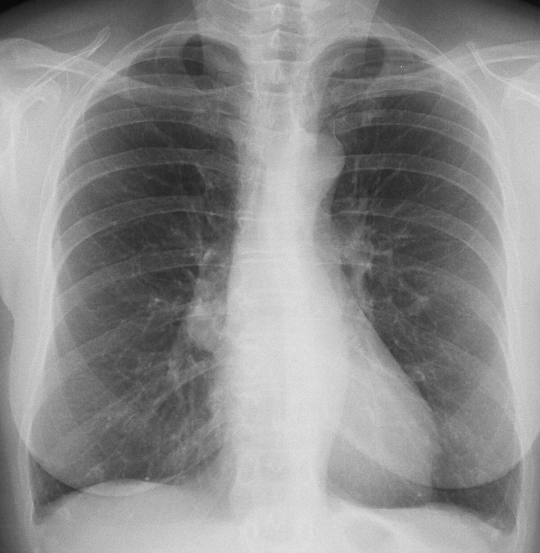
Today I am showing the second case of the “five easy pieces”. It is a PA radiograph of a 51-year-old woman for pre-op surgery of umbilical hernia. What do you see?
Check the image below, leave me your thoughts in the comments section, and come back on Friday for the answer.
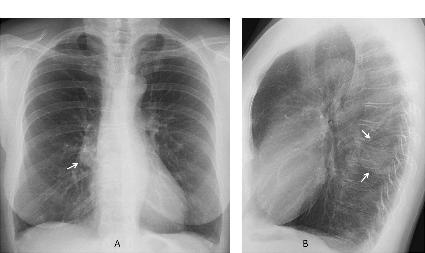
Findings: PA chest radiograph shows increased opacity of the lower right hilum (A, arrow). Lateral view was taken and demonstrated that the opacity was located posteriorly (B, arrow) and had features of an extrapulmonary lesion.
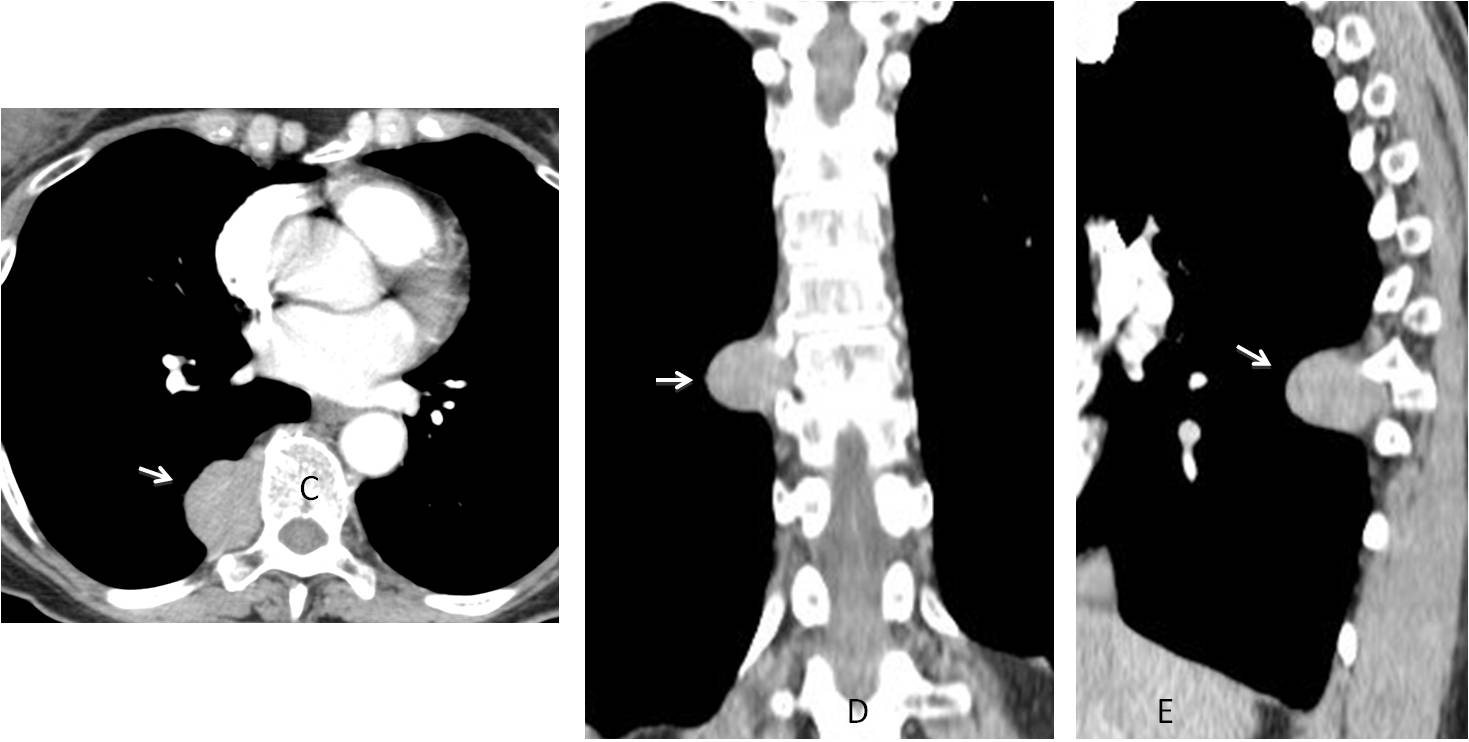
Enhanced CT confirms a solid posterior mediastinal mass (C-E, arrows). The most likely diagnosis is a neurogenic tumour. At the time of writing, surgery is still pending.
Final diagnosis: posterior mediastinal mass simulating an enlarged hilum.
Congratulations to all of you (many) who saw the abnormality, with special mention to Olena, who gave the first answer.
Teaching point: the most common cause of prominent hilum is lymph nodes or an enlarged pulmonary artery. Occasionally it may be simulated by superimposition of a mass located in front of or behind the hilum, as in this case.


There is additional homogeneous round opacity in the right hila – needs lateral veiw
Differential diagnosis – bronchopulmonary cyst with fluid ? mediastinal mass?
The shadow of aortic deformation from aortic arch to the level of diaphram, which deforms the carina – it shifts carina to the right
Rt hilar mass
honeycomb on the left overview for interstitial lung disease? + Minimal pleural effusion = scleroderma?
right mediastinal lap sarcoidosis ?
Hello,
Opacification of left-lower part of trachea and round opacity in a right hilum. I suspect npl of bronchi with lymphadenopathy.
Paracardiac oval shaped opacity below right hilum, without silhouette sign, therefore probably situated posteriorly – bronchogenic cyst is my first suspect.
Slightly blunted costophrenic sulci, probably of no significance.
CP angles blunted.Right hilum full.
Aortic shadow within normal limits.
Junction line visible superior aspect aortic knuckle.
Any back pain?
Round opacity, below the right hilum, probably situated posteriorly – lung cancer?
Opacity of right hilum.
Aneurysm of descending aorta with right hilar opacity. Lymph node more likely.
Descendan thoracic sırta aneurism and right hilar opacity( Lap?)
Right parahiliar adenopathy
Blunted phrenicocostal sinuses due to pleural effusion. Fusiformly dilated descending aorta. Homogenous round opacity in right hilum, possibly adenopathy.
…senza una LL non ci si può sbilanciare…..potrebbe essere un esito adenopatico come da m. di Castleman…aspettiamo la TC del venerdì….un caro saluto da Bari….
Any right pleuritic chest pain
No. What were you thinking?
(I apologise prof.Jose i wos wery busy -i’m on duty in regional hospital) suspect for discret air -fluid level ( a littl amount of the likid) right subdiafragmal.Anyway i will do a chest x ray in expiration.
On the other hand radiography is asimetrical,therefore no mediastinal structures are entirely clear – x ray anatomy.
Solitary pulmonary nodul and blunted phcostal sinuses – my opinion is granuloma.
Chest x ray in full expirium -susp. a small c.phrenic pneumotorax dex.
No neumothorax. Sorry
I got it! Buy my English is not as good as I though. I tried to post a report in English, buy I couldn´t find the words (or maybe I was concerned that I wrote it bad). Can I use Spanish in a next case?
You can write in spanish, of course. I need to practice!
Right hilarious mass
Tracheal shadow not continuous
Scleroderma??
You haven’t confirmed or denied pleural effusions. Could you clear that up for us? Love your posts. Thank you!