Showing today pre-op radiographs for a hip prosthesis in a 62-year-old man.
What do you see?
Check the images below, leave your thoughts in the comments section, and come back on Friday for the answer.
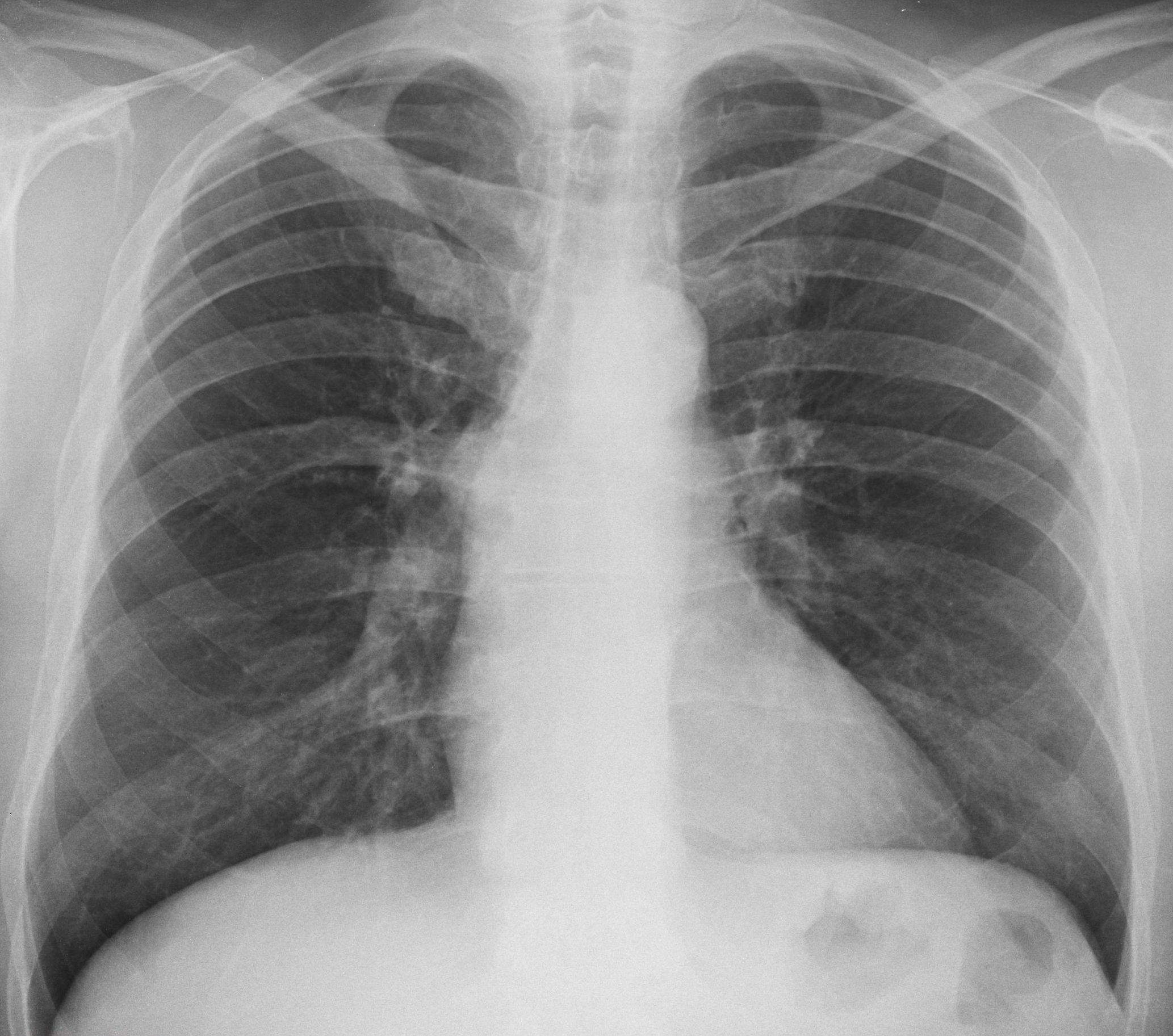
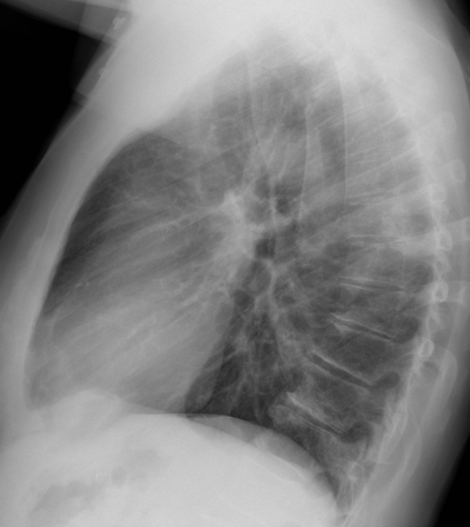
Findings: PA radiograph shows an extrapulmonary lesion projected over the right hilum (A, arrow), located in the posterior mediastinum in the lateral view (B, arrow).
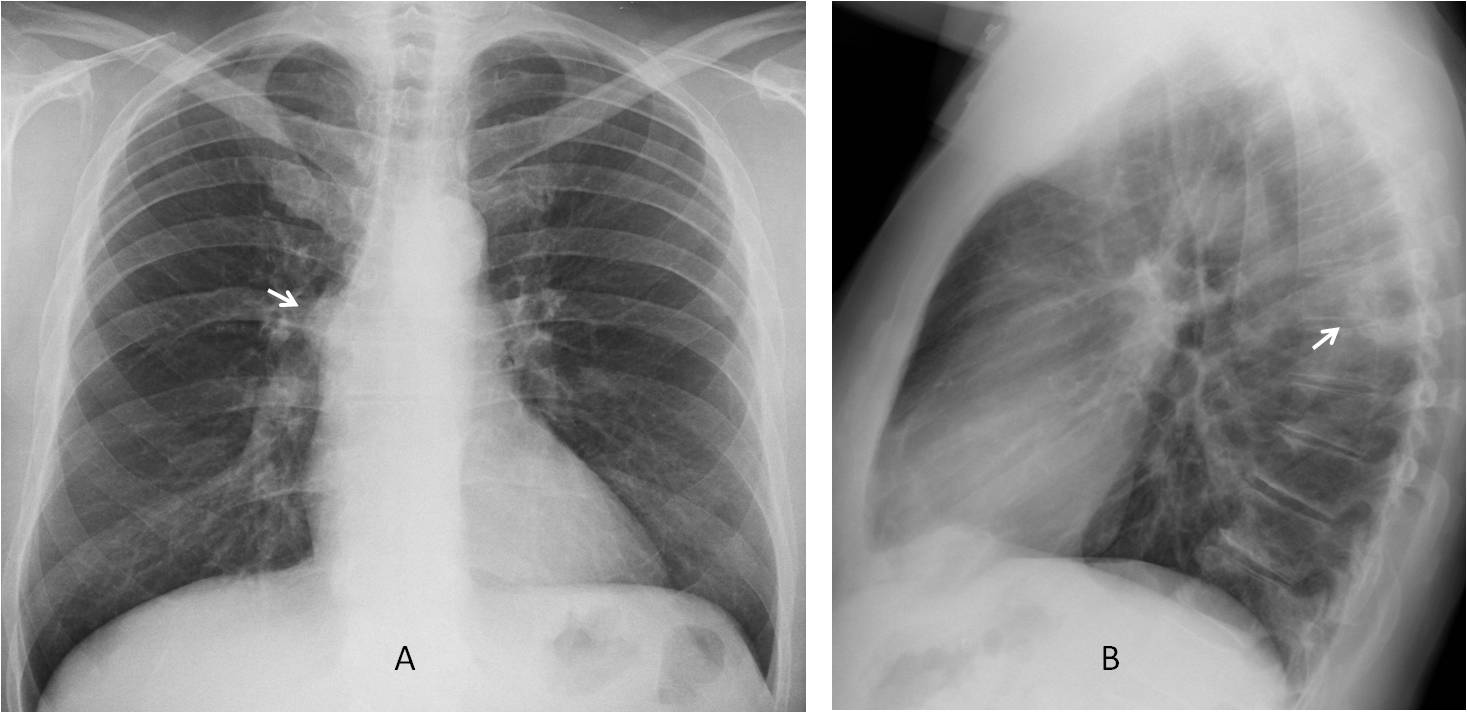
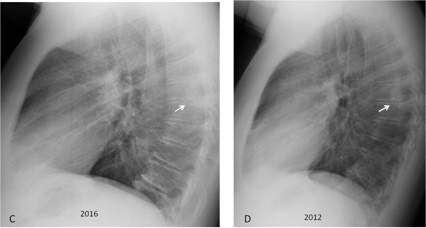
The lesion is very similar to the one shown in case 135 and neurogenic tumour is the first diagnosis that comes to mind. Comparison with previous film shows that the lesion has not changed in a four-year interval (C-D, arrows).
Coronal and sagittal CT demonstrates that the lesion corresponds to a large osteophyte (E-F, arrows).
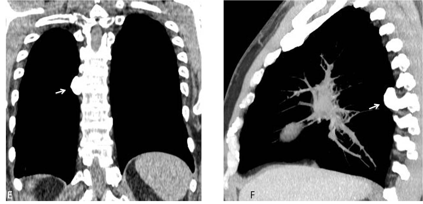
Final diagnosis: osteophyte simulating a posterior mediastinal mass.
Congratulations to all of you who saw the mediastinal lesion. It is important to emphasize that high KV makes it difficult to recognise calcium.
Teaching point: remember that not all posterior mediastinal masses represent neurogenic tumours. Processes arising from the spine and mediastinal fat should also be considered (see Dr. Pepe’s Diploma Casebook case 64).


I noticed a thick walled cavity at the posterior LLL which is more obvious on the profile X-ray. Additionally, there is a fracture/ lytic lesion on the Rt posterior 8th rib. Finally, there is some hyperinflation..
Some pneumosclerosis, such as age-related
First day, no answers. Are you getting cold feet?
right hilar rounded opacity
There seems to be increased radiodensity of the posterior elements of two middle thoracic vertebrae. Maybe metastatic lesions?
Subpulmonic effusion on the left?
Cavitary lesion best seen posteriorly on lateral view. Apparently overshadowed by the right hilum on frontal view.
Looks thick-walled, worrisome for neoplasm (bronchogenic carcinoma, metastatic). Ddx includes infeccion (tuberculosis, fungal), vasculitis and rheumatoid nodule.
I think an extrapulmonary posterior mediastinal cavitary mass would unsual.
Do you see it in the PA view?
Maybe I’m seeing too much. I thought it was projected over the R hilum at level of the 8th posterior rib.
And maybe what I thought was a central cavity on the lateral view is just the neural foramen.
Your interpretation is correct. Final diagnosis tomorrow.
Two opacities seen on lateral- one projected over upper thoracic vertebra and one over lower thoracic vertebra. Neither are cavitating. Only one is seen on PA view projected over upper right hilum.
They look quite dense but are not obviously calcified
Given that there are two metastases would be the most common diagnosis.
CT would be next step.
Metastases are no the only cause of vertebral opacities.
rt. thoracic spinal neurofibroma?
Sensible diagnosis
….professore stimatissimo…..ci sono 2 elementi da considerare, tutto a dx, a livello di D8: esiti di rimaneggiamento costale posteriore dx ed opacità a livello del corrispettivo forame di coniugazione intervertebrale….esiti di neurinoma ?….
Any alternative diagnosis to neurogenic tumor?
….forse un linfangioma ….
SAPHO syndrome – a long shot and probably wrong!
Like your diagnosis, although it’s not the right one. Answer tomorrow.
Some form of benign hyperostosis of the vertebra. Doesn’t look typical for DISH. I am at the limits of my MSK knowledge.
Close enough. You get full credit for the answer, although it is too late to include your name in today’s answer.
Well, if this is really a vertebral sclerotic lesion I’d put melorheostosis in ddx list.
wait for the next case!!!
Is there any litic lesion or old fracture in the 8th right rib?