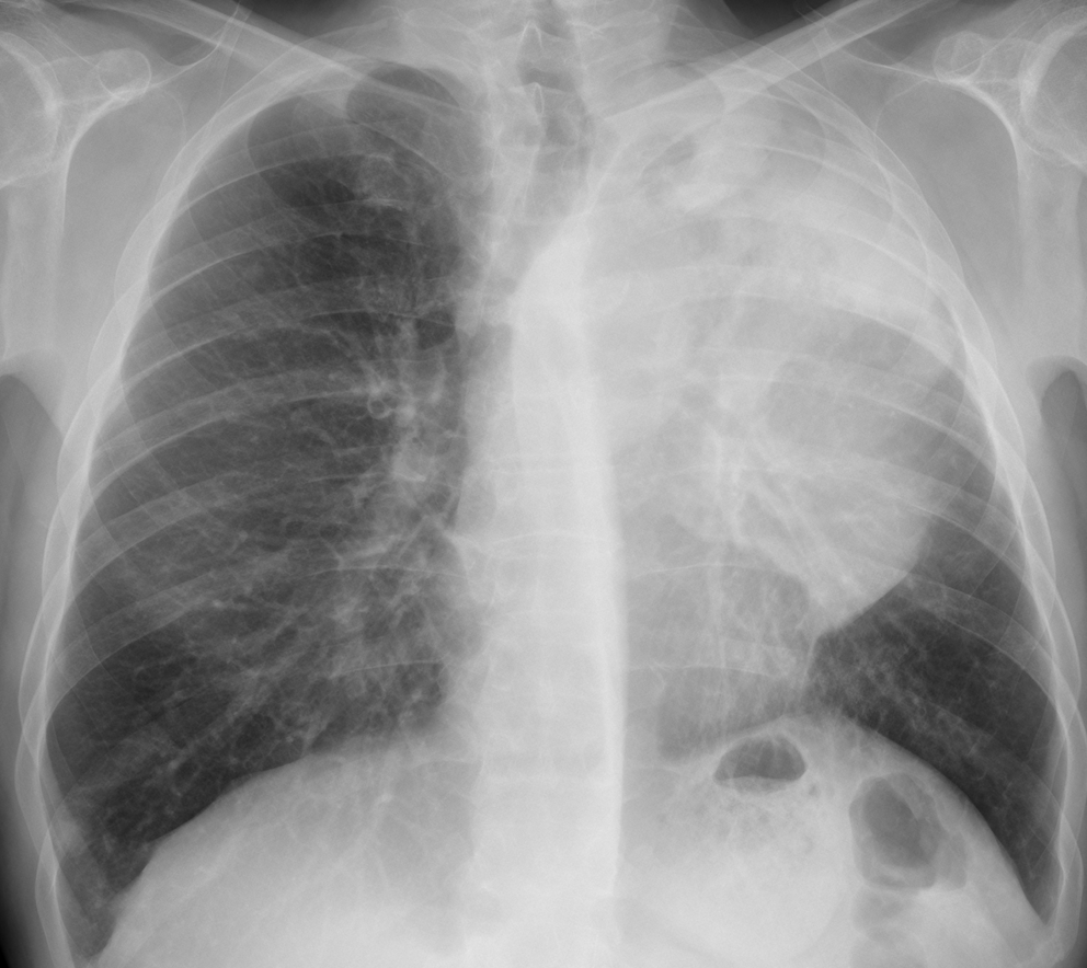
Today we’re showing chest radiographs of a 57-year-old man with chest pain and dyspnoea.
Check the images below, leave your thoughts in the comments, and come back on Friday for the answer.
1. Carcinoma of the lung
2. Thymoma
3. Lymphoma
4. None of the above
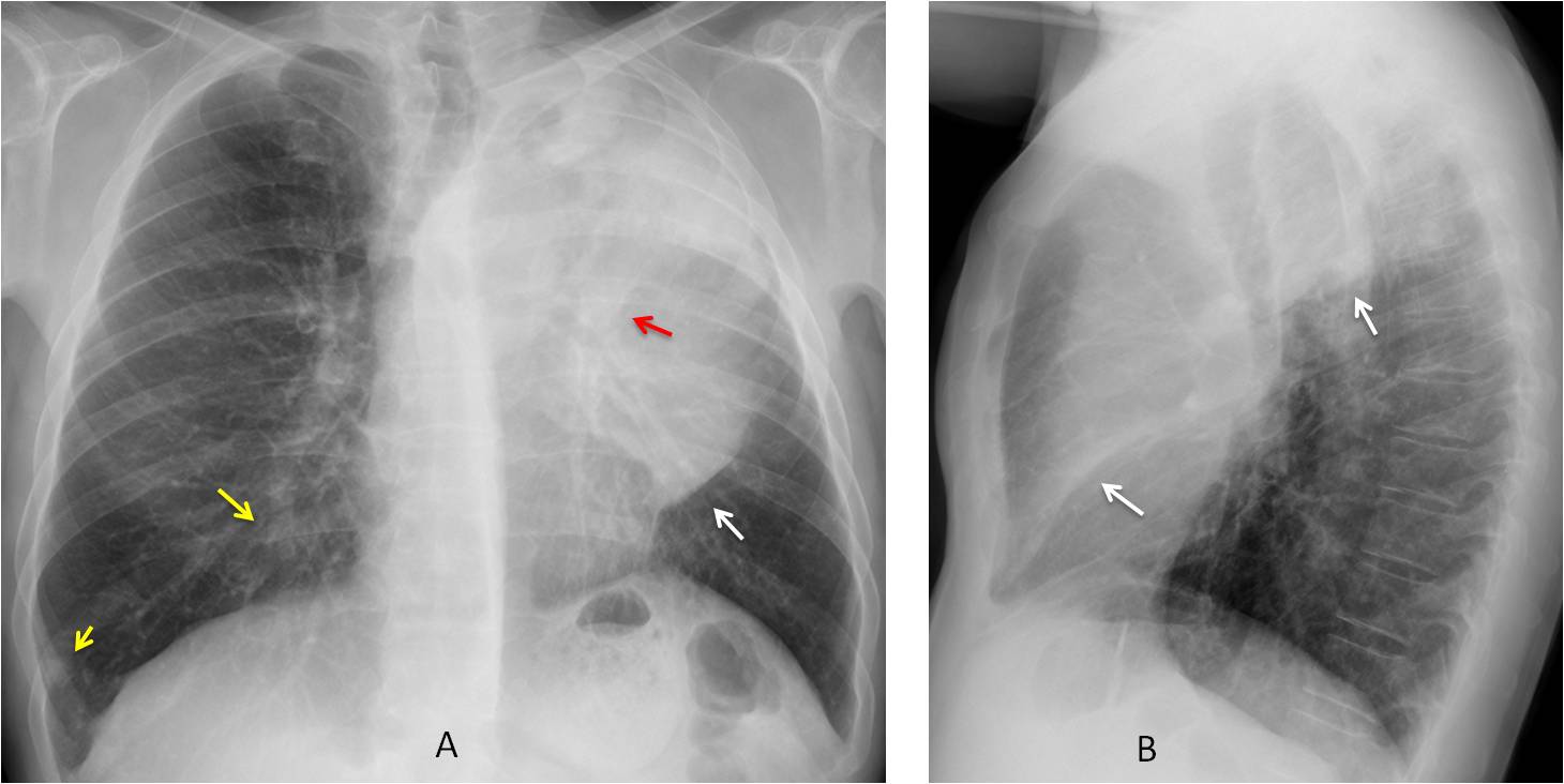
Findings: the PA radiograph shows opacity in the left lung, partially well defined inferiorly (A, arrow). The trachea is displaced and the left hilum is slightly elevated (A, red arrow). This appearance is highly suggestive of LUL collapse with a central mass. Additional findings are several nodules in the left lung (A, yellow arrows), which, in this context, are most probably metastases.
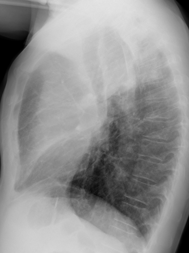
The diagnosis is reinforced by the lateral view, which shows the typical pancake shape of the collapsed LUL, limited by the major fissure (B, arrows).
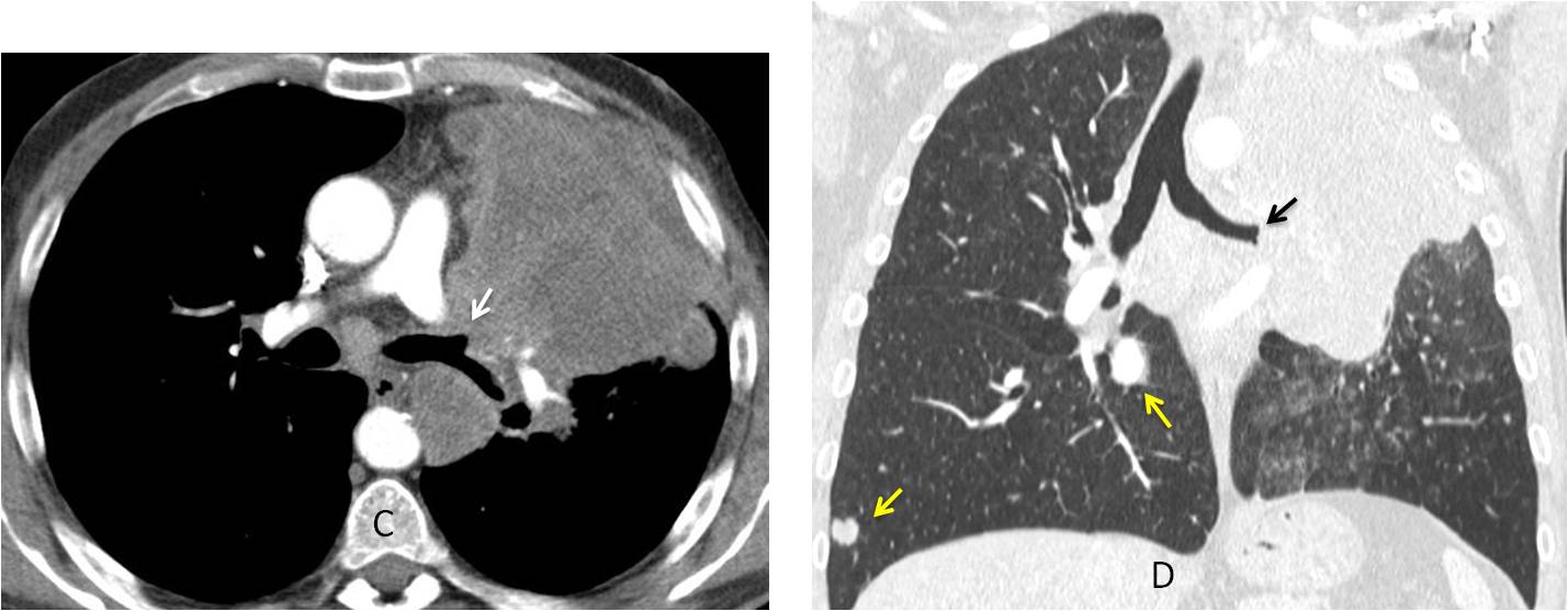
Enhanced axial and coronal CT confirms the LUL collapse and occlusion of the LUL bronchus (C-D, arrows). Nodular metastases are visible in the right lung (C-D, yellow arrows).
Final diagnosis: carcinoma with LUL collapse and metastases to right lung
Congratulations to MK, who was the first to make the right diagnosis.
Teaching point: diagnosing LUL collapse is reasonably easy for a competent radiologist. Seeing the right lung metastases takes you to the first division. Remember to avoid satisfaction of search!


There is a superior tracheal shift towards the left side, but it´s inferior portion is displaced towards the contrateral side.
In the lateral view there is a partially obliterated retrosternal space.
I think there are signs of atelectasia of the LUL, well delimitated for the major fissure, and I think that a Golden´s sign is visible.
The left bronchious is elevated and the cardiac silhouette is missing.
I think that the correct answer is a carcinoma of the lung with hiliar/mediastinal adenopathy.
Giant aortic arch aneurysm?
Mr. Marian…stai cuminte
On the PA radiograph there is an oval mass adjacent to the mediastinum on the left, causing displacement of the distal trachea to the right. On the lateral view this mass appears to be in the middle portion of the mediastinum. Additionally on the PA, there is opacification of the left hemithorax with visible bronchogams, as well as silhouetting of the heart and the medial portion of the left hemidiaphragm. On the lateral view there seems to be a wedge-shaped density overlaying the cardiac silhouette along the anterior portion of the left major fissure. These findings may represent a thymoma with an associated collapse of the left upper lobe.
Interlobar pleural effusion!!!
1-There is volume loss in left upper lobe evidenced by superior displacement of major fissure on lateral view, horizontal orientation of left main bronchus on PA view and leftward displacement of trachea in PA view. Finding are suggestive of left upper lobe collapse
2- the obvious mass in PA view turns to be in middle mediastinum in lateral view and the retrosternal air space on lateral view appear enough lucent to exclude thymoma and lymphoma as a cause of this large opacity.
Finally, my diagnosis is carcinoma of the lung complicated by left upper lobe collapse.
carcinoma of the upper lobe .
colapso del lobulo superior izquuierdo por un cancer del pulmon
By the way, did anybody look at the right lung?
Perhaps at least 2 pulmonary nodules? One in the URL and the other in the LRL
Why didn’t you see them? Do some soul searching
We didn’t see because of satisfaction syndrome
Good. You learned a lesson.
Thanks, professor.
….bulking linfoma con atelettasia lobo polmonare superiore sx.
thymoma