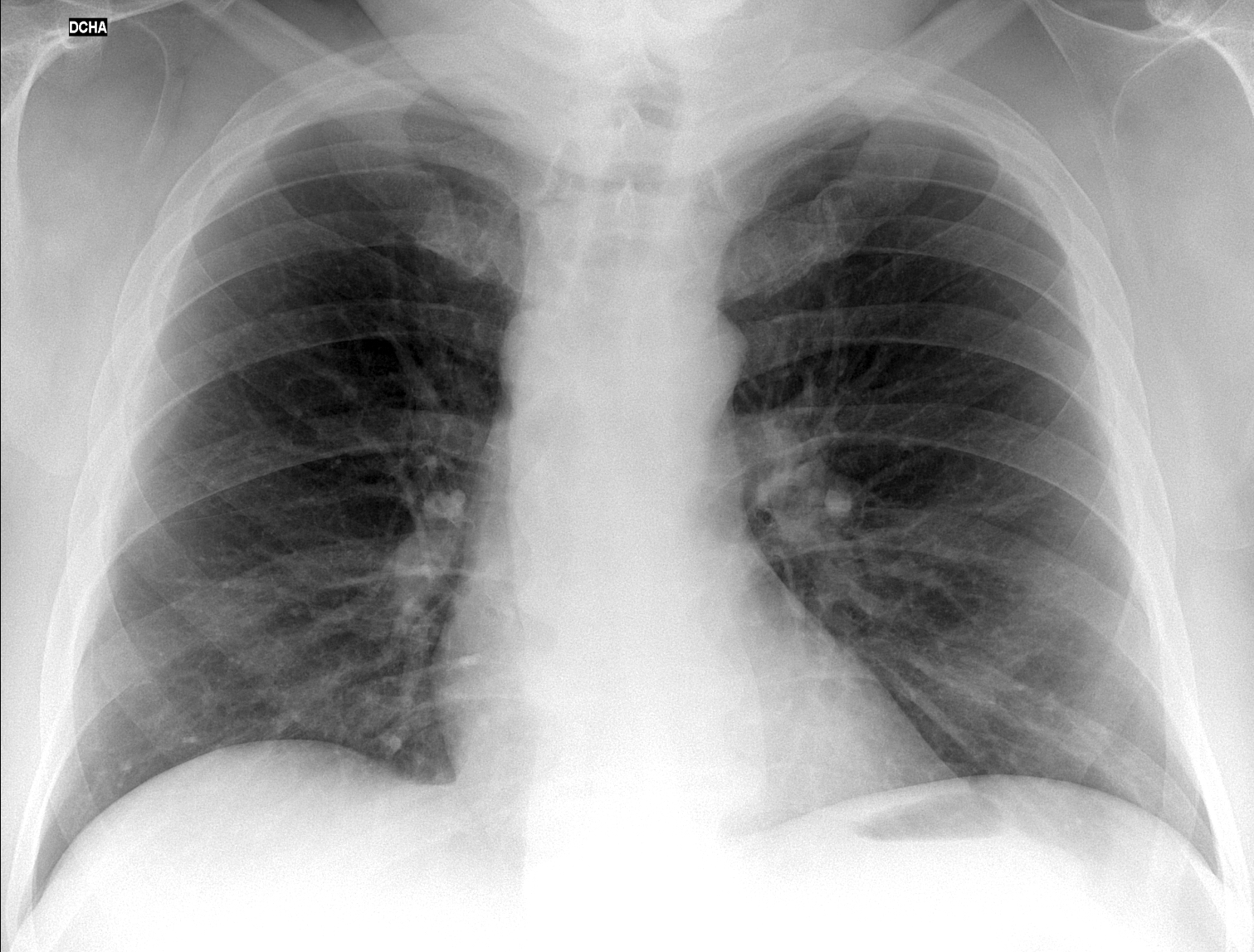
Today we are presenting a pre-op PA chest radiograph of a patient with inguinal hernia. What do you see?
Check the image below, leave your thoughts in the comments section and come back on Friday for the answer.
Findings: the PA radiograph shows a rounded shadow in the right upper mediastinum (A, arrow). All mediastinal masses look the same in the plain film; but the clue to the diagnosis lies in the presence of big osteophytes on the right and left sides of the spine (A, red arrows), suggesting that the rounded shadow also represents a big osteophyte. A lateral view taken two years ago shows osteophytes at the same level (B, arrow), accompanied by calcification of the anterior ligament.
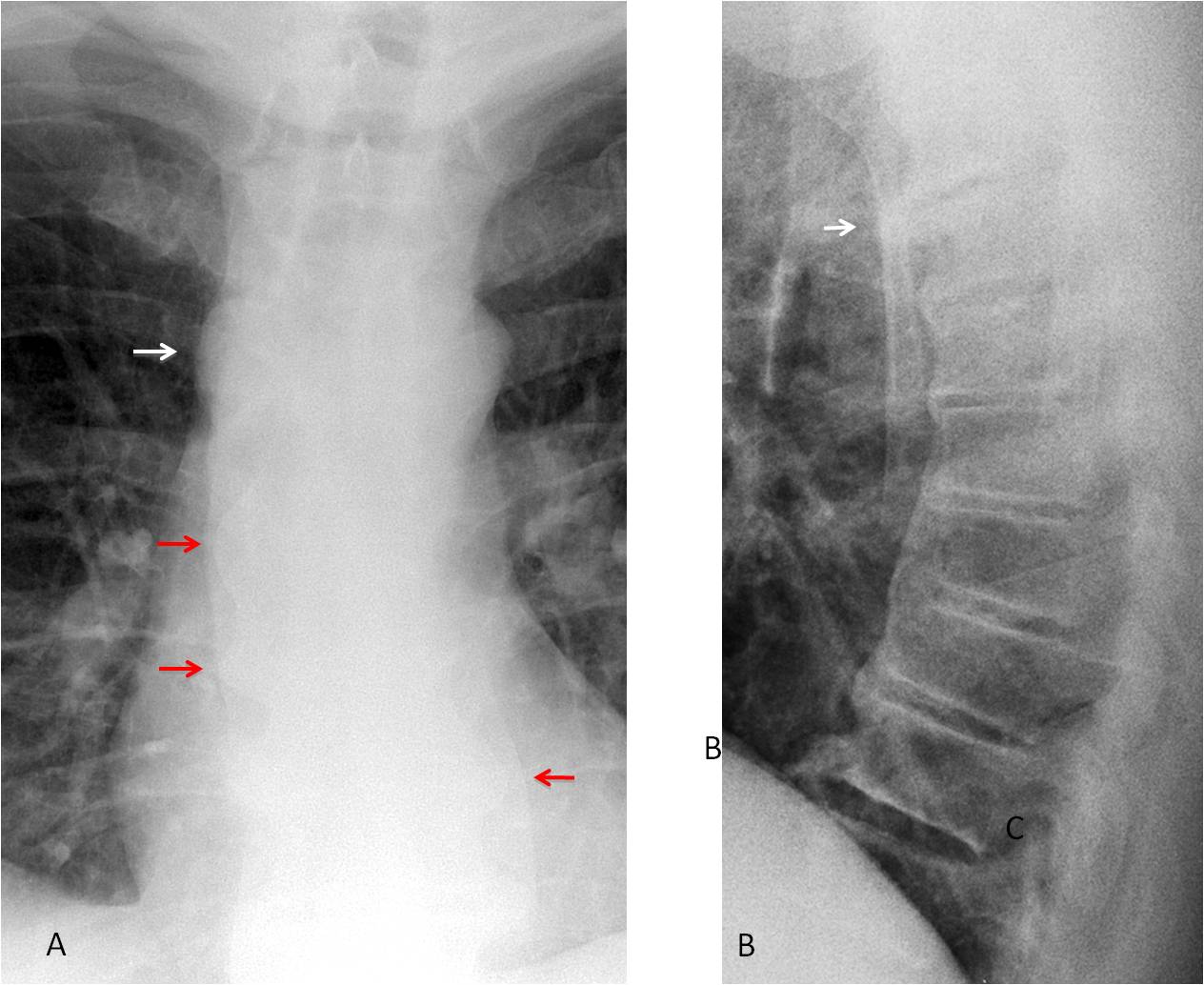
Axial CT shows that the mass corresponds to a large osteophyte (C, arrow).
3-D reconstruction shows extensive calcification that seems to flow along the anterior aspect of the spine (D-E). This appearance suggest a possible DISH
(Forestier disease).
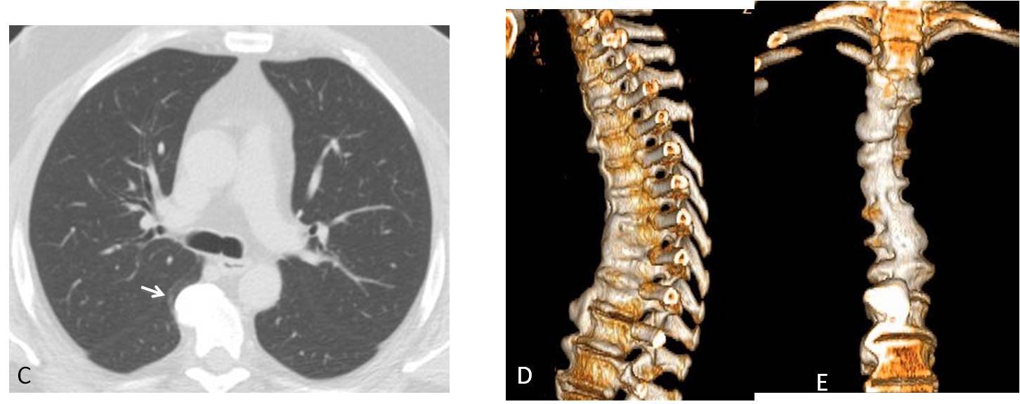
Final diagnosis: osteophyte simulating a mediastinal mass in a patient with probable DISH (unproven).
Congratulations to ZHW, who was the first to suggest the diagnosis.
Teaching point: do not forget that a large osteophyte may simulate a posterior mediastinal mass. And in this patient led to a probable diagnosis of systemic bone disease.


Hello.
There is a small area of bulging of the right upper mediastinal contour. Double aortic arch?
Hello! I think that the bulging of the right mediastinal contour is because of the azygos arch. So, we can see a displaced right paraspinal interfaces, because of osteophytes.
It is a poor inspirated PA x-ray. The right hilum is lower that the left one.
lytic lesion of the right scapula
….professore facendo nostra la tua precedente lezione ….dilatata la vena azygos…..le cause , di questa supplenza emodinamica, le hai elencate nella lezione precedente del dr Pepe….ilBari finalmente VINCE….il Barca ancora non conVINCE….
….una alternativa potrebbe essere un piccolo aneurisma del l’arco aortico…
Hmmm…there is pleura changes on the both sides, wide v.azygos, pulmonary art. hypertension? IX rib destruction on the left?
Ribs are OK. I would like to see prominence of the pulmonart arch before suggesting pulmonary hypertension
Am seeing this on phone, so maybe missing a lot of things.
Right paratracheal bulge – ?rt sided aortic arch/ ?prominenr azygos
iPad gives much better definition
Bilateral paraspinal line widening due to ostrophytes
Thickening along bilateral hemithoraces subpleural .this is likely due to subpleural fat.other consideration could be pleural fluid or thickenimg.
What do you think of the bulge of the upper right mediastinum?
Hmmm… wide v.azygos, pulmonary arterial hypertension? IX rib destrukcion on the left?
Ups..sorry
One of you described findings that may help to suggest the etiology of the bulge in the upper mediastinum
Perhaps the right paraspinal line (tubular structure) is the margin of the continuation azygos because of the interruption of the inferior vein cava
The right-sided bulging of the mediastinum at the level of the aortic knob looks like continuation of the azygos vein with interruption of the inferior vena cava. A contrast enhanced CT is needed to confirm.
http://learningradiology.com/archives2011/COW%20466
Azygous%20Contin/azycorrect.htm
http://learningradiology.com/archives2011/COW%20466Azygous%20Contin/azycorrect.htm
I guess the link won’t paste properly. Sorry…
Hello Professor,
– Well defined focal para tracheal bulge on right side
– Prominent right para spinal line
consistent with continuation of azygous vein with interruption of the IVC
Confirm with CT chest and abdomen with IV contrast
smooth focal right mediastinal bulge at the level of the aortic knuckle.
Features are likely to represent prominent azygos vein with differential being double aortic arch or lymphadenopathy.
So far you all have mentioned as the ethiology of the bulge: right aortic arch, aneurysm, enlarged azygos. You haven’t mentioned lymph nodes or other mediastinal masses. It is none of the above. You are missing a clue.
The down right hilum can be because of mass effect?
No. As a matter of fact, I do not think that the hilum is low
Ok
Associated with the bulging I think there is widening of the space between the ribs. Maybe a posterior mediastinum neural lesion? Schwanomma?
Other consideration would be a pleural lesion. Fibroma?
No, sorry. Answer tomorrow
The right paratracheal lesion interupts and causes apparent displacment of the right paravertebral line so is in the posterior mediastinum. Given all the osteophytes elsewhere it could just be more of the same
Bingo!!
artic ectasia
ectasia of the aorta
what the meaning picture on your blog?