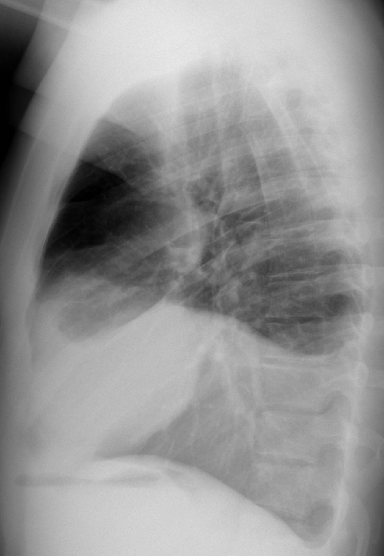
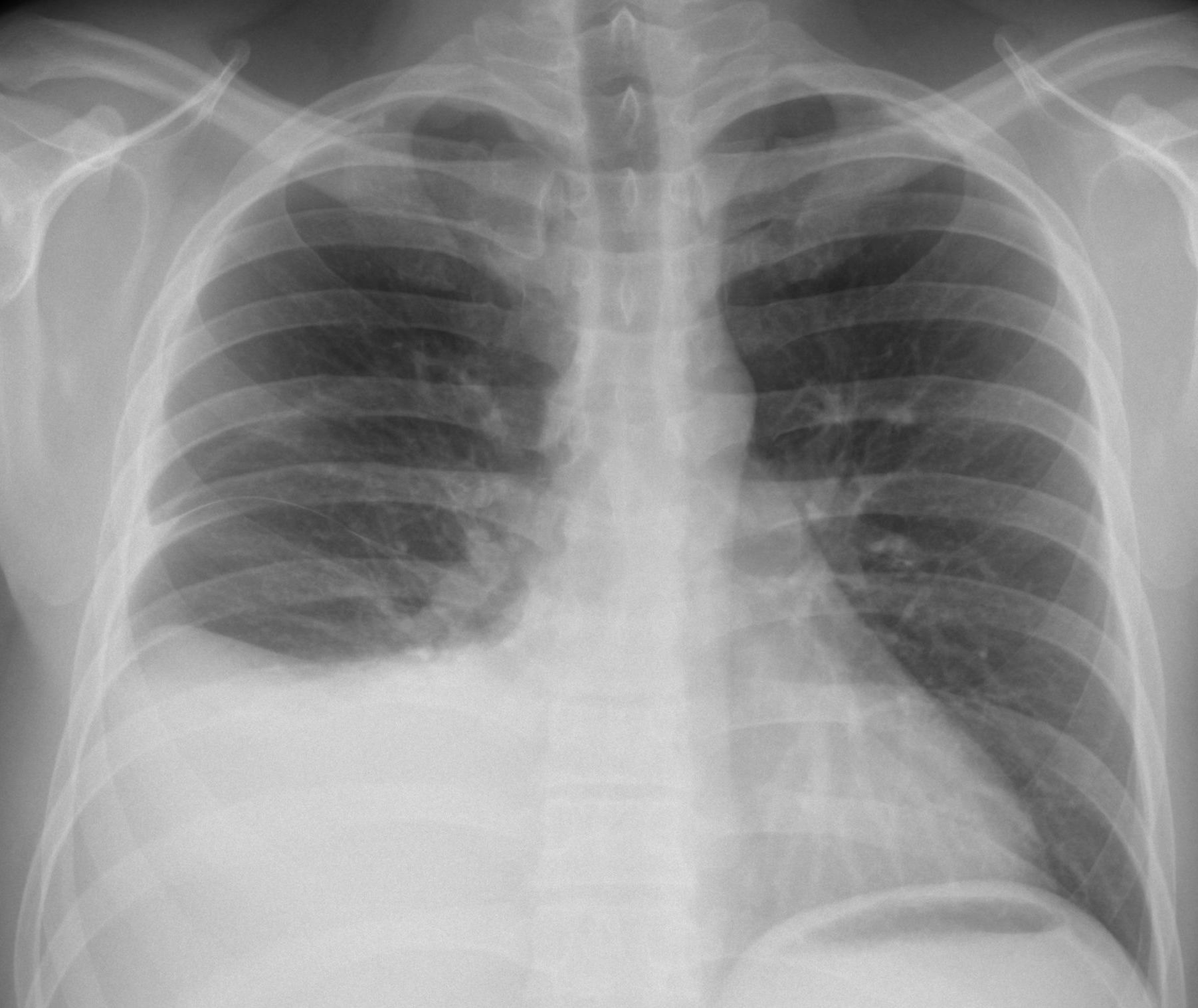
Today we are presenting radiographs and CT images of a 35-year-old male tourist from Venezuela, who came to the ER with pain in the right hemithorax for the last five days. No fever.
As usual, check the images below, leave your thoughts in the comments section and come back on Friday for the answer.
1. Mesothelioma
2. TB
3. Metastases
4. Any of the above
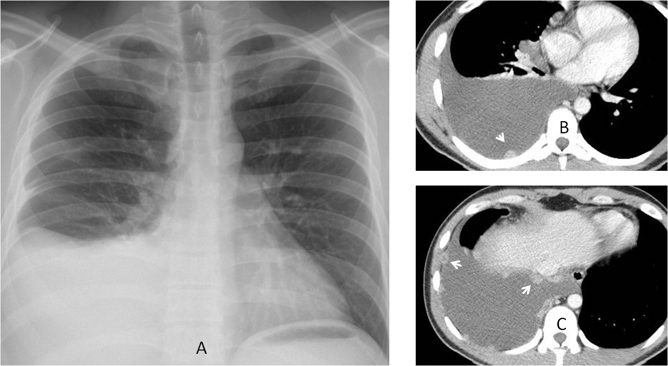
Findings: the chest radiograph (A) shows a right pleural effusion. Axial CT shows numerous enhancing pleural nodules (B-C, arrows).
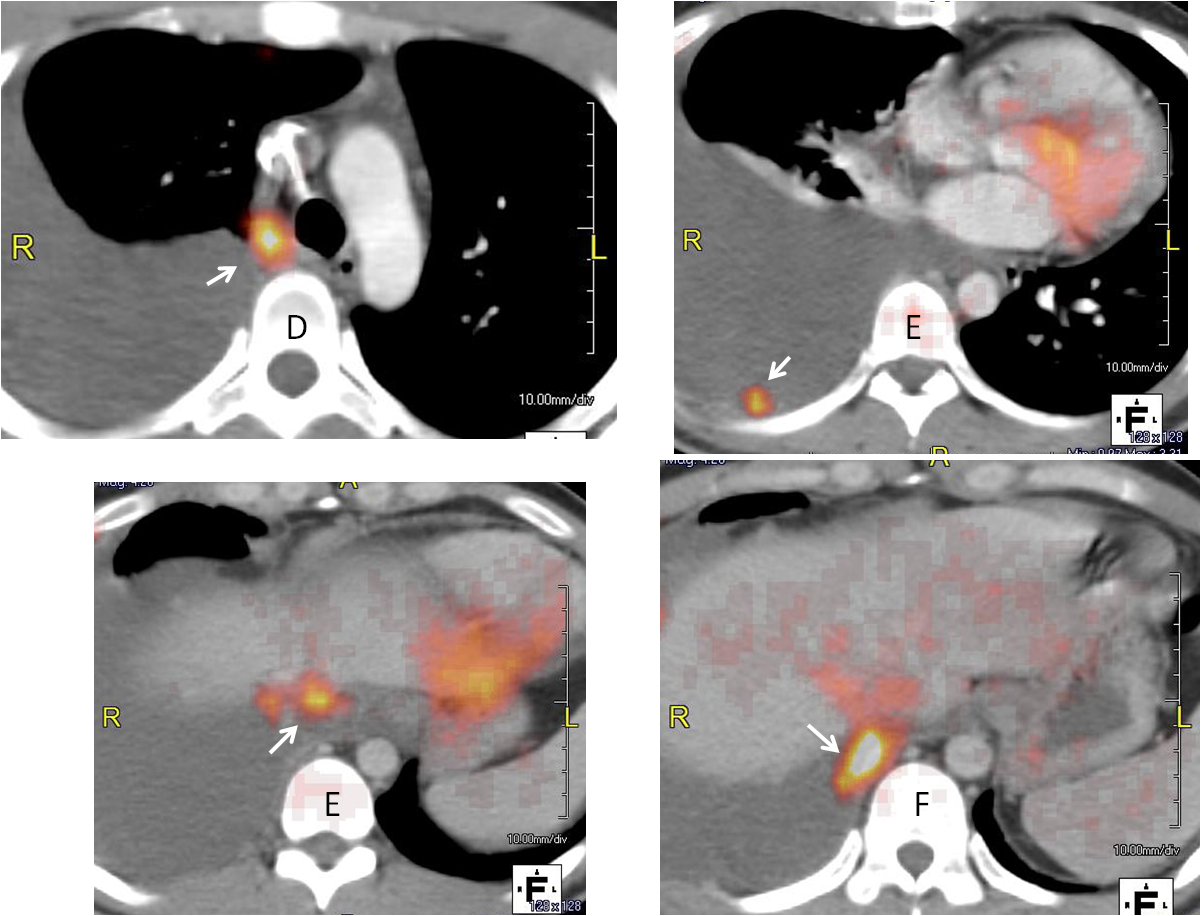
PET-CT shows uptake of the nodules (D-G, arrows)
When I saw this case, I thought of malignancy as the first possibility (either metastases or lymphoma). To my surprise and shame, pleural biopsy revealed casseifying granulomas, BK positive. I did not consider TB because I had never seen pleural nodules in TB and there was no lung involvement.
I wanted to share this case with you because it is a first for me, although this presentation of pleural TB has been described. I am not surprised about the positive PET-CT, because I have seen it many times in active TB.
Final diagnosis: tuberculous pleuritis.
Congratulations to Genchi Bari, who made the diagnosis in unequivocal terms.
Teaching point: remember that TB is a great imitator. It should be included in the differential diagnosis of malignancy of the chest.


Hi,
right pleural effusion with multiple enhancing pleural nodules.
no suggestive signs of TB.
the age and appearance don`t suggest mesothelioma, although can`t be excluded.
in this age and with this appearance i think the probable diagnosis is metastatic disease (osteosarcoma?..).
thanks
Hi,
a hypodense lesion in the right hilum, seen in the lower CT image, might be the primary source of metastases.
There is right pleural effusion with nodular pleural lesions along the mediastinal and costal pleura. Thickening of the right minor fissure noted without nodularity.The morphology of the lesion is not very specific for malignancy and in a young patient with fever infection cannot be excluded. So I think any of the above could be possible. Pleural fluid analysis and biopsy should be suggested. Thank you.
Hello!
Right pleural effusion with nodular mediatinal and parietal pleural hypervascular lesions, some of them with hypovascular center (mediastinal nodular pleural lesions tends to be
Malignant).
The air that is under the left diaphgram… is it on the Stomach?
Mtx from a carcinoid perhaps?
In the PA x-ray there are some apical extrapulmonary lesions and in the right paratracheal margin, probably more pleural lesions.
Any of above
Metástasis pleurales
….Prof. …..TBC pleurica…..i noduli captano mdc ed un nodulo presenta un centro ipodenso , come da necrosi caseosa.
Good diagnosis. Congratulations for Juve’s victory!
…..grazie mitico blu-grana……ricorda Dybala può’ essere il futuro vincente del tuo Barca !!!!!
Hello,
-Right pleural effusion + enhancing nodular pleural thickening.
Possible diagnosis: pleural tumor (mesothelioma, lymphoma), metastases.
Right massive pleural effusion with multiple nodules localized on visceral pleura of the posterior and right lateral chest wall, on the diaphragmal pleura and also on mediastinal pleura, growing in pericardium (?) some of them have smooth well-defined and other on the contrary ill-defined and irregular contours, and also some of them have small region of hypointensity
the additional mass on CT in the right hilum
Really very nice and comlicated case. Only one-sided type of changes, chest pain but no fever make to suggest firstly malignancy/mts. however if to suggest tuberculosis – it is not typical for such tuberculosis changes to be without fever, only with chest pain.
The anamnesis is also a pitfall – Venezuela it is exotic country so any other rare diseases can differentiated(parazites, infections)
If to take in consideration the x-ray images some peribronchial changes are seen predominantly in midddle zones and on the right and pleural plaques are also seen on the right apex.
nice case…