This weeks’ radiographs belong to a 31-year-old male with vague chest complaints. What do you see?
Check the images below, leave your thoughts in the comments section and come back on Friday for the answer.
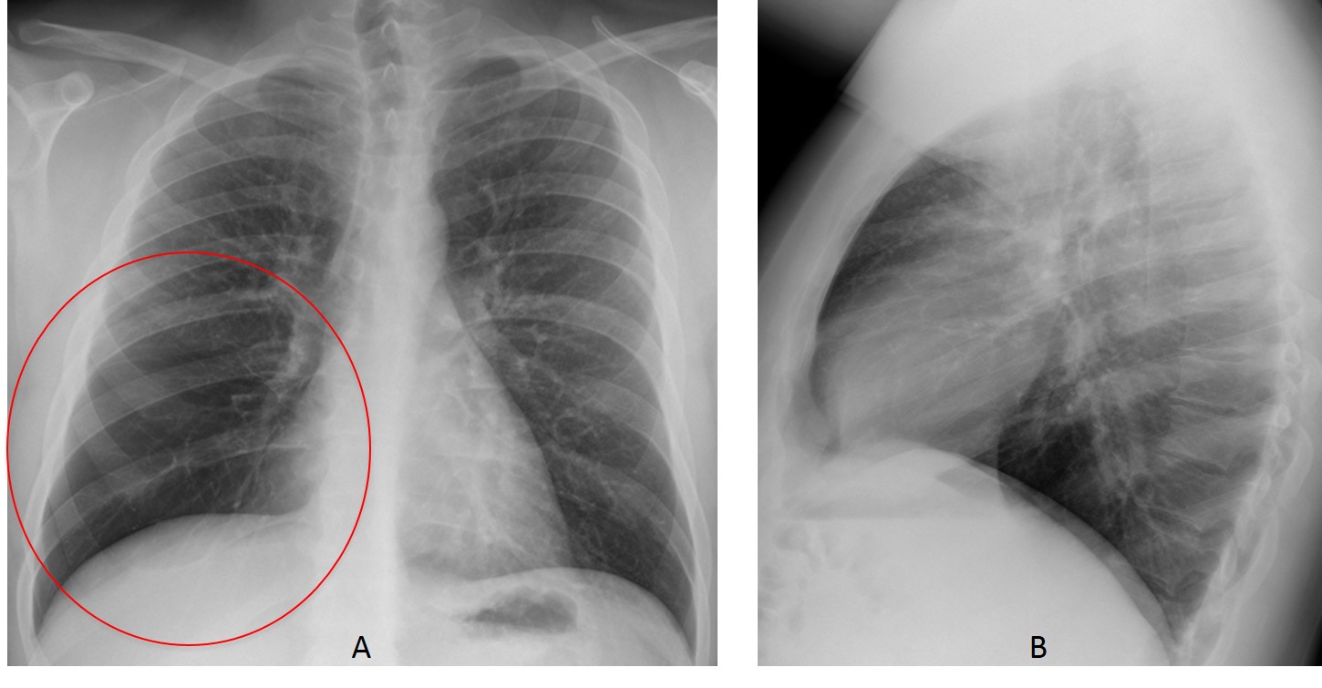
Findings: The lateral view is unremarkable. PA radiograph shows increased lucency of the lower right lung with decreased vasculature (A, circle).
This finding has two main causes: increased lung air or paucity of lung vessels (pulmonary embolism, arterial stenosis). In these cases, the best approach is to take an expiratory film, which will demonstrate whether or not there is air-trapping. If present, it will orient us to an obstructive process, either central or peripheral.
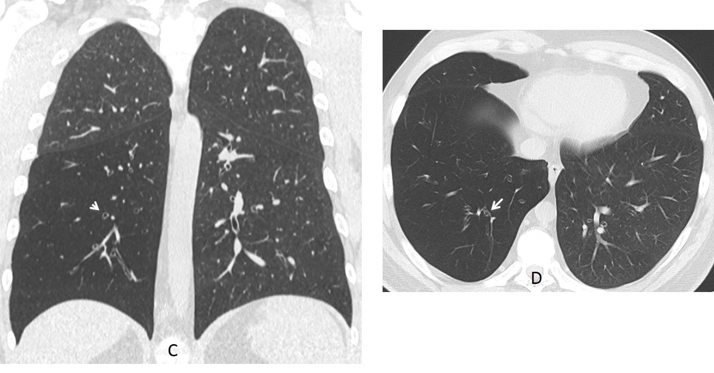
Unenhanced CT confirms the increased lucency and diminished vasculature of RLL and RML. In addition, scattered bronchiectasis are seen (C-D, arrows).
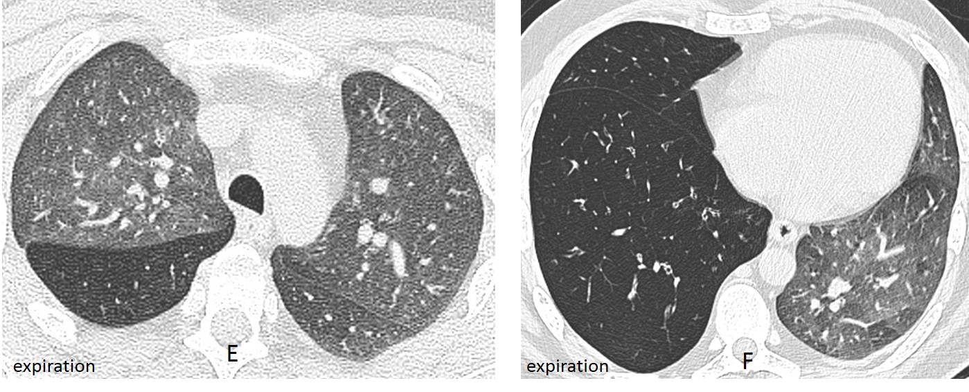
Axial expiratory CTs (E-F) demonstrates air-trapping.
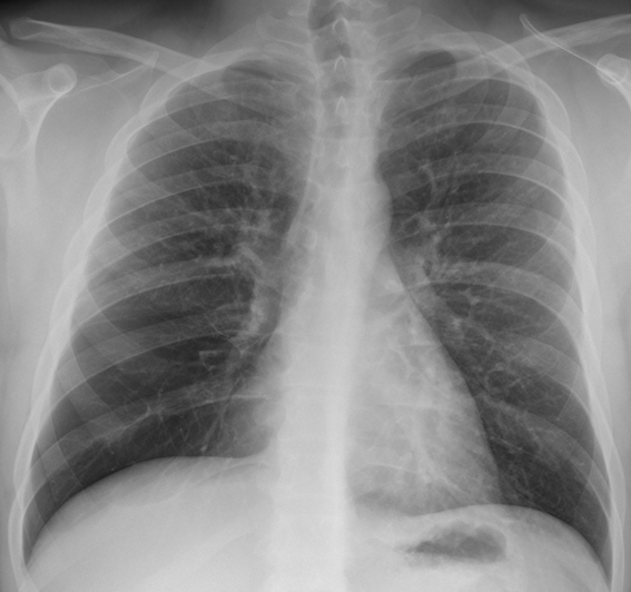
The patient had a history of swallowing a peanut at the age of five years, developing RLL pneumonia at that time. Control radiographs demonstrated increased lucency of the lower right lung over the years.
Final diagnosis: Lobar Swyer-James/McLeod syndrome secondary to aspiration of a peanut in childhood.
Congratulations to TR, who was the first to submit a firm diagnosis.
Teaching point: remember that expiratory films in hyperlucent lung are very useful to orient the differential diagnosis (see Dr. Pepe’s Diploma Casebook case 86)

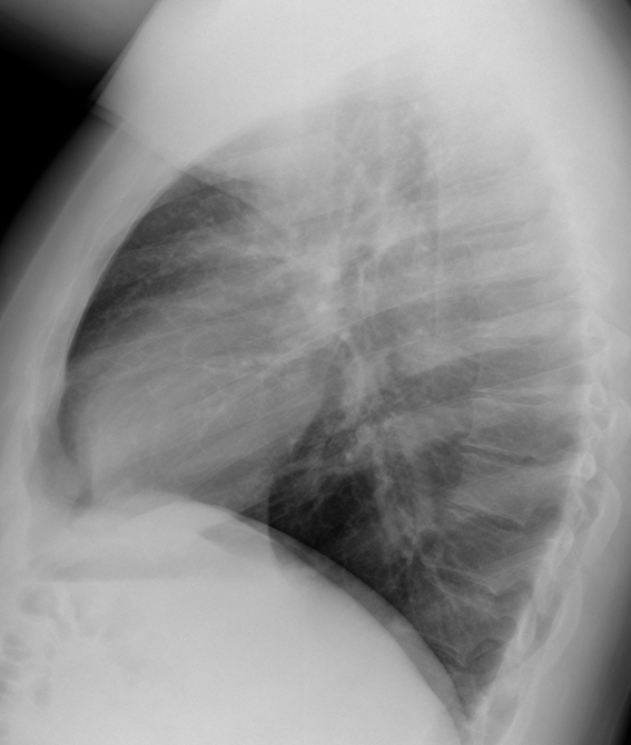


Good morning! The x-ray is rotated, so the right paratracheal line is thickened.
There is an increased density proyected over the left cardiac region, that in the lateral view has vascular appearance. The lower right hemithorax has less vasculature than the left one.
The right cardiac silohuette is not well define and in the lateral view there is a vertical line, so pulmonary hypoplasia is a good idea…
Define pulmonary hypoplasia
It is an incomplete developtmen of parts of the lung.
Perhaps the paucity of vascular markings is becuase of MacLeod syndrome?
I would like to demonstrate air-trapping with an expiratory film.
Perhaps subtle findings to think on pulmonary hypoplasia….so not good idea
Patient’s position is rotated due to the skoliosis, that is why the projection of the mediastium, trachea seems abnormal
There is hyperlucent RLL with further poor lung vasculature, making decrease attenuation of the upper part of diaphragm – bulla of the RLL
The changes first suggesting bullous disease
But the absence of thin lines caused be thin walls of bulla, is against this. As the absence of pneumothorax as common and often complication too.
Maybe there is a vascular abnormality – vascular hypoplasia/ or lung hypoplasia.
Righ middle lobe aplasia
What would you do to prove this diagnosis?
I looked over radiographs and my previous comments and would like to re-formulate my thoughts:
1. I think unilateral pulmonary hypoplasia can be excluded – usually it is associated with scimitar syndrome, other vascular abnormalities; affected lung is small, mediastinum shifted into the ipsilateral hemithorax.
2.Next two abnormalities should be differentiated – agenesia and aplasia, the last one has a rudimentary bronchus.
These abnormalities can affect both lung and one lobe isolated.
On this radiographs it is more likely isolated lobe agenesia/aplasia because there is compensatory hyperexpansion of the RLL and as a result distortion of vessels.
Though there is compensatory hyperexpansion but still the lung volume is reduced (mediastinum slighly shifted to the right and elavated diaphragm)
There are one more two abnormalities for differentiation: bronchial atresia (common triad – central mucocele, hyperlucency and hypoperfusion of affected segment) and congenital lobar overinflation (neonatal manifestation)
The best way to prove this diagnosis is to do bronchoscopy
9th rib from right side is abnormal
cardiac bronchus
Hello,
I’m agree with Olena – there is less vasculature of lower pole of right lung. Conclusion – Westemark sign in PE.
The right heart border is poorly defined on the PA. The vessels extending from right hilum seem crowded and displaced medially.
On the lateral I see a faint opacity overlying the heart. To me this seems wedge-shaped, and I also see vascular structures within the retrocardiac clear space.
Although my knowledge is limited, I think this is a right middle lobe collapse with compensatory hyperinflation of inferior right lobe.
I would do CT to confirm and rule out retrocardiac or endobronchial mass.
Thank you for letting me participate.
Greetings,
hypelucency of the right lower lung zone with diminished vascularity, no increase or decrease in volume, no enlargement of the right pulmonary artery, my suggestion is Swyer-James syndrome/obliterative bronchiolitis.
There is hyperlucency of the right lower lung, with segmentary hypovascularization. Was this Xray taken in ER? Pulmonary embolism?
Patient was practically asymptomatic
ipoplasia arteria polmonare dx, con ipolasia POLMONARE, MI SEMBRA UNA BUONA DIAGNOSI…..ASPETTANDO L’ANGIO-TC….DA bari un caro saluto….
scoliosis upper thoracic spine .
hyper ventelation rt lower lobe lung