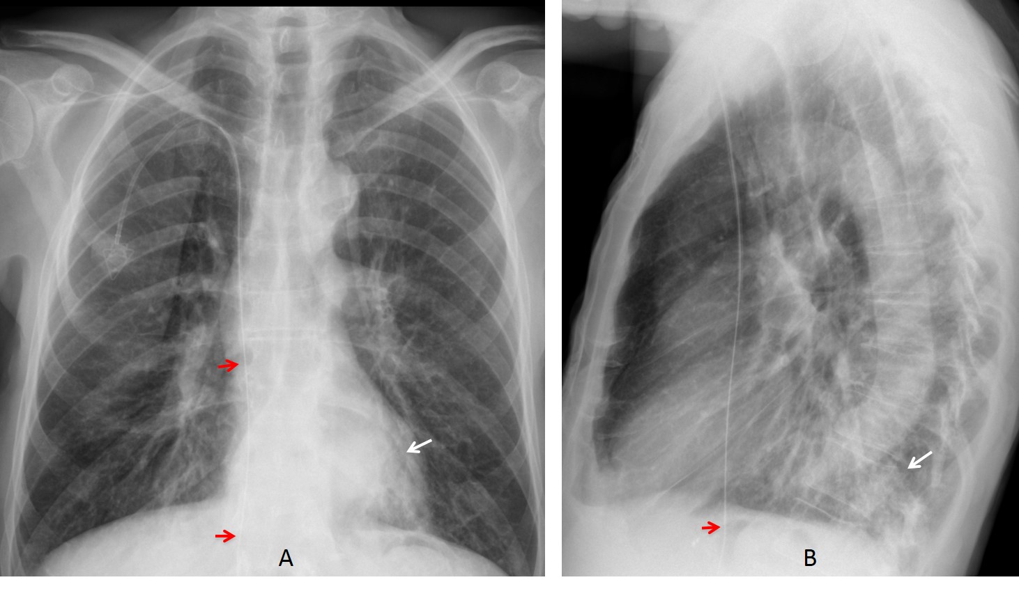
Case 1 findings: PA chest radiograph shows a retrocardiac pulmonary infiltrate (A, arrow), confirmed in the lateral view (B, arrow). Although the patient did not have significant symptoms, it was considered to be inflammatory and cleared with antibiotics. An additional finding is the path of the guiding wire of the port-a-cath, which is clearly going beyond the right atrium into the abdomen (A-B, red arrows).
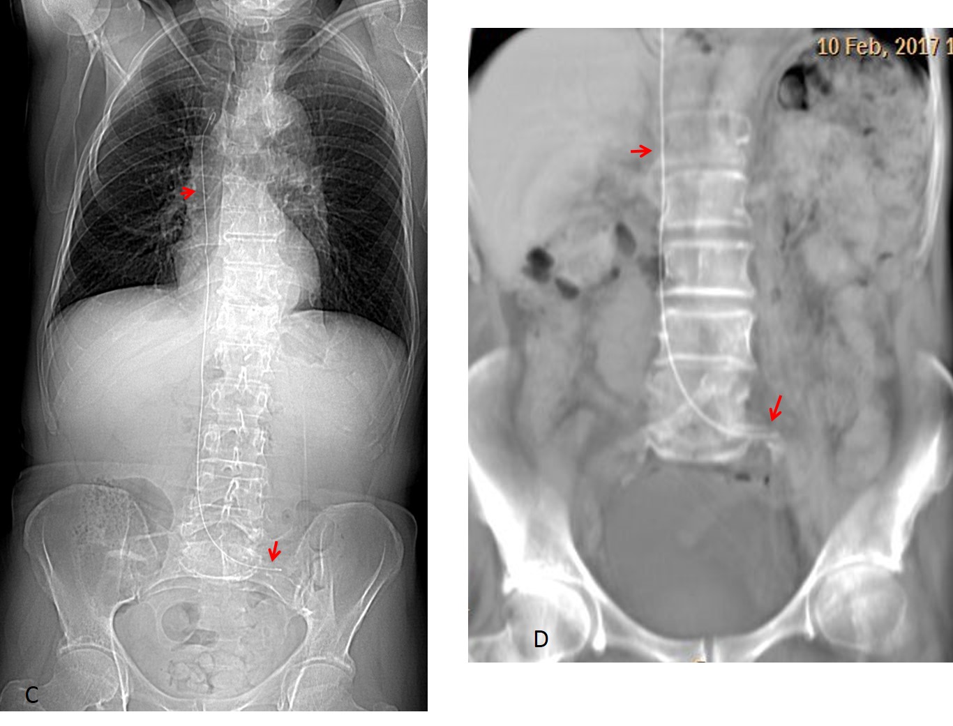
CT scout view after removal of the port-a-cath shows the wire remaining in the SVC, with the distal end in the left iliac vein (C, arrows). Coronal CT taken four years later shows no change (D, arrows).
Final diagnosis: LL pneumonia. Wire lodged in SVC and IVC.
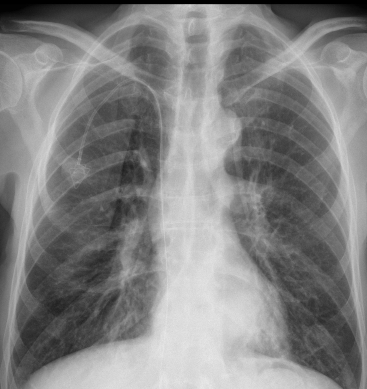
Case 2 findings: PA chest radiograph shows fibrotic changes in the right apex and a faint nodule in the mid left lung fiend (A, arrow), not visible one year earlier (B, circle), raising the possibility of a metastatic nodule.

Axial CT and 3-D reconstruction show that the nodule represents a healed fracture of the anterior fifth rib (C-D, arrows) accompanied by another fracture of the sixth rib (D, yellow arrow).

Final diagnosis: unsuspected rib fracture simulating a metastatic nodule.
Congratulations to LAR, who saw the abnormal path of the port-a-cath and to Coffee, who was the first to detect the nodule if the left lung.
Teaching point: I selected these two cases to cover for Dr. Pepe, thinking that the diagnosis would be easy. Remember the KISS method!


Good morning!
In the first case there is a retrocardiac left opacity (I would like to see the lateral view) and an aereal paramediastinal vertical line.
The right cardiac sillouhette is missing, probably because of postsurgical changes.
In the second case there is a right retrocardiac opacity but I think it is the left atrium (double contour sign). In the right apex there are probably fibrotic changes. Right breast surgical changes.
The early bird does not always get the worm!
See the answer tomorrow
In the first case, there is focal mass-like opacity at retrocardiac region, further lateral view is helpful.
In the second case, there is suspected increased density at periphery of left mid lung, possibly along left anterior ribs, quesionable for bony lesion. There are reticulation at right upper lung, possibly post infection or post radiation
I mostly agree with your assessment, with some additions:
In case #1 I would also mention a lucency that exists in right middle zone medially, but I’m not sure of the origin.
In case #2 there are reticulo-nodular markings in middle and lower zones bilaterally, suspicious of lymphangitis carcinomatosa. There is also some scaring in upper zones, but non-significant.
Perhaps I am deceived, but I see a soft tissue mass in stomach in case #1.
It is probably food. No mass was present on CT.
Tueasday night and only two comments. Cases are either too easy or too difficult!
Yes, these are difficult but very educational 🙂
In image 1, something is causing loss of the right heart border. There also seems to be increased density of the pulmonary parenchyma in this region. I would consider fibrotic changes post radiotherapy, middle lobe collapse, or less likely, pectus excavatum. There is also a left retrocardiac opacity as mentioned by MK and Coffee.
In image 2, there seem to be fibrotic changes in the right apex. I also think the mediastinum is widened on the right. Could be a widened paratracheal stripe due to mediastinal lymphadenopathy.
CT should be done in both cases given their history of cancer.
Hope both cases are educational 😉
Greetings,
Case 1: large opacity seen through the left heart shadow. suspected round opacity over the right middle zone obscured by the external end of the central line. tortuous azygoesophageal line.
Case 2: faint peripheral opacity over the left middle zone. possible fibrotic changes over the right apex.
Greetings after a while,
In the first picture there’s a lot of diverson caused by vertical linear opacities probably caused by skin folds or sth. similar. As mentioned above theres seems to be a quite large retrocardiac rounded/nodular opacity also the tip of the central line/vascuport is placed too low.
In the second picure all the suspected reticular or fibrotic changes are very faint and to me of no clinical meaning. What’s interesting I think I see right sided cervical rib.
Regards Professore..
Case nr 2.
LINEAR FIBROTIC CHANGES IN RIGHT APEX WITH CALCIFICATIONS. BENIGN.
BOTH LUNGS WITH MULTIPLE SMALL NODULES PROBABLY METASTASES .
ON THE LEFT OPACIFICATON ALONG THE ANTERIOR PART OF IV RIB -ALSO META SUSP.
case nr 2.
Opacification on the right is probably a swab covering a needle inside iv acces.
Patient position asymetric.
Retrocardiac and perivertebral opacification metastatic lymphadenopathy
First case: perm-a-Cath too low, in IVC.
Right lateral deviation of the azygoesophageal line with ill defined retrocardiac mass, tumor recurrence, lymphadenopathy? Obscurantion of the right heart border with depression of the right hilum, right Middle lobe collapse. Review of lateral X-ray is required. The shadow of the right scapula is increased?
Second case: right axillary surgical clips with abnormal contour of right breast, due previous surgery. Fibrotic changes in right superior mediastinum, post radiation? Right retrocardiac opacity, lymphadenopathy? Cortical defect in the right scapula laterally with focal lucencies, metastasis? Review lateral and bone scan for further assessment
….1 rx …..la punta del cvc è’ fuori posizione e in vcs. 2 l’arco anteriore della 5 costa sx è’ Mal visibile per probabile riassorbimento Litico….ho conquistato i 3 punti.?
You are close. Will give you one and a half points 😉
See answer tomorrow