As promised, here are the two additional images. Do they help you to reach a conclusion?
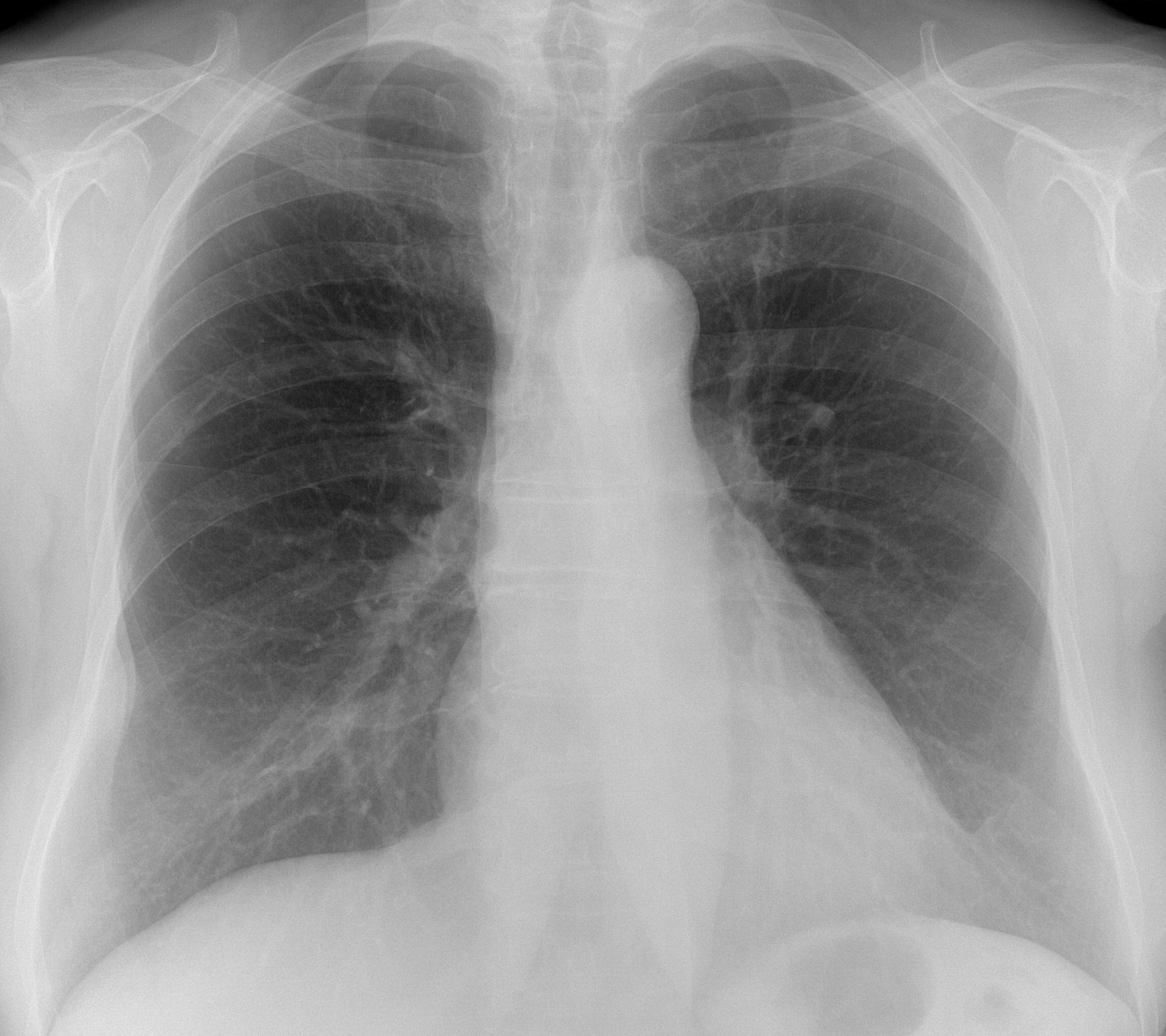
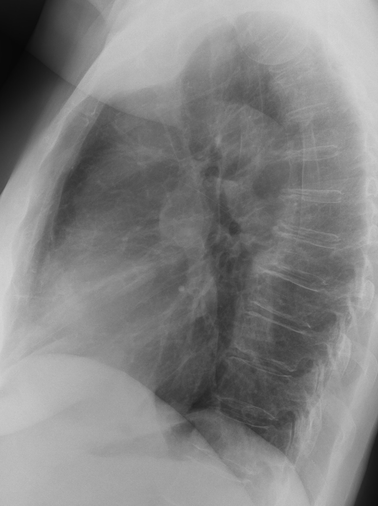
Findings: the PA radiograph shows mild compression of the right wall of the cervical trachea due to a known goiter. The main finding is the presence of an extrapulmonary lesion in the lower right hemithorax (A, arrow) with possible enlargement of the underlying rib giving a double contour to the lesion (B, arrow).

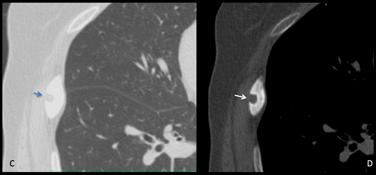
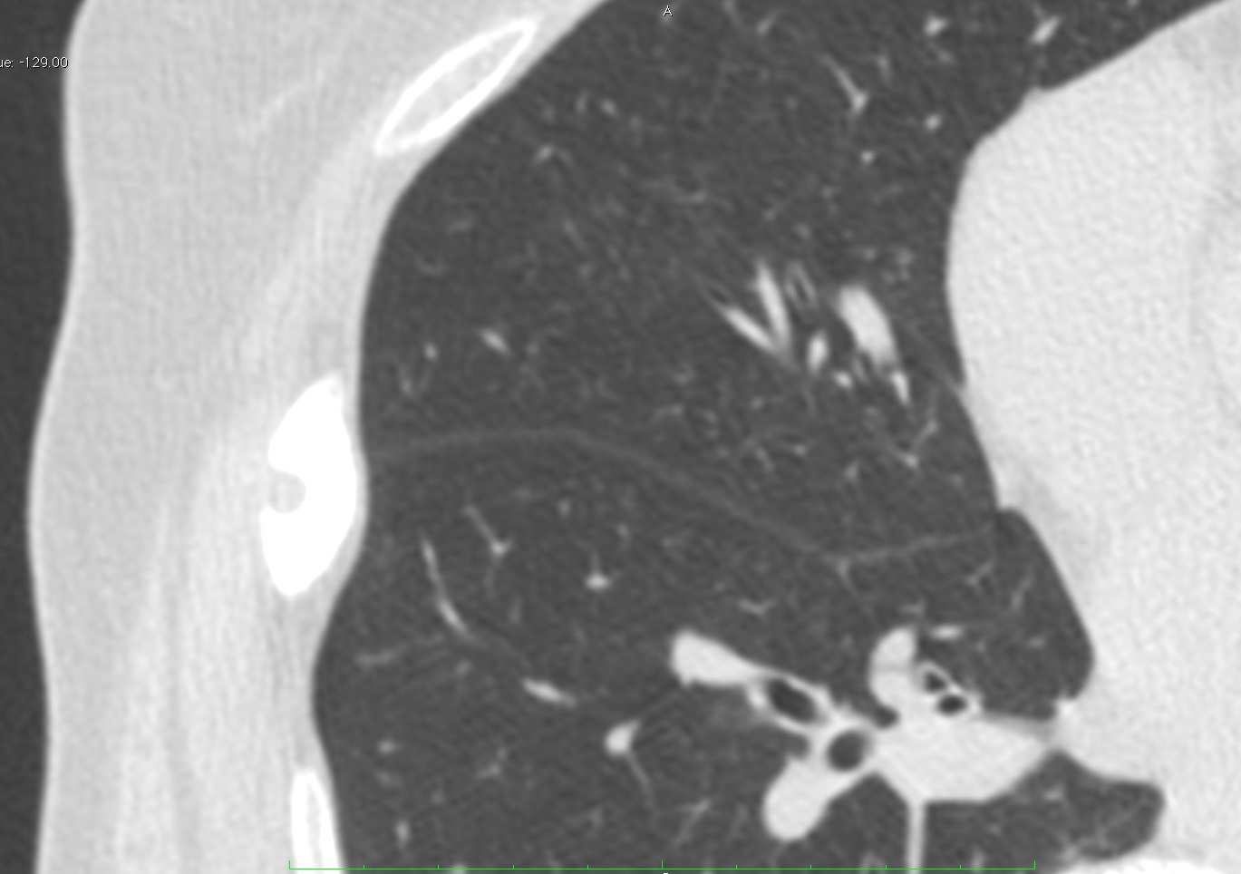
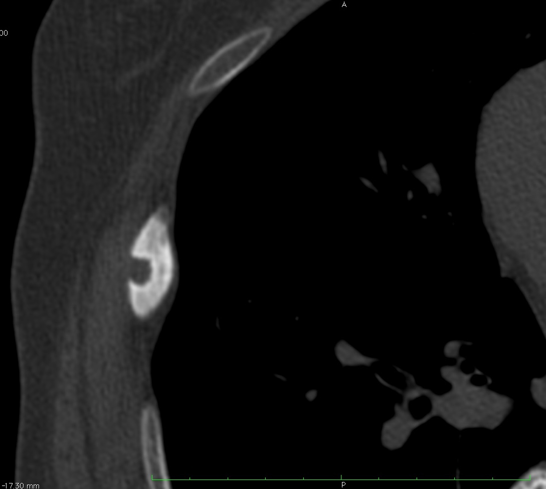
Unenhanced axial CT shows a widened sclerotic rib with a well-defined punched-out lesion in the outer cortex (C-D, arrows) with sclerotic border.
As I mentioned when presenting the case, I don’t have a definitive diagnosis. The lesion is clearly slow growing, as evidenced by the sclerotic border. Certainly it is not an osteoid osteoma (no pain and no swelling of soft tissues). The peripheral location and the aspect go against enchondroma. I cannot rule out fibrous dysplasia, a common lesion in the rib, but it does not look like one to me.
After interrogation, the patient recalled a previous fall while in the country many years ago, hurting her right side. My explanation is that she got a sharp vegetal foreign body in the rib at that time, which ended in a foreign body rib granuloma.
The patient is asymptomatic and I will do a CT follow-up in six months; will share the results with you here on the ESR blog.
Tentative diagnosis: Foreign body granuloma of the rib (unproven)
It is difficult to congratulate anyone, since I don’t have the right answer. In my opinion, Sadaf and Coffee were the people who better analysed the findings.


Good morning!!!
There is a pleural lesion at right hemithorax, for example an hematoma because of costal fracture (any hystory of trauma?).
In the lateral x-ray the right hilum has a particular round shape, but I cant see any anomaly in the AP proyection.
It was a routine examination and trauma was not mentioned in the petition
Fibrous pleural tumor?
…..guarda la trachea , in AP e LL, a livello della biforcazione…..impronta e stenosi ab -estrinseco…..
2 finding
Pathologische Pleura thickening rightside in level of lowerfeld.
Thickening of the right paratracheal line with deviation of trachea to liftside.
DD Pleural carcinomatosis
Extrapulmonary lesion laterally on the right, it might as well be a schwannoma, although I do not see any additional finding supporting this diagnosis.
Saludos desde Hungría, gracias.
Wait for CT tomorrow 😉
Hello. I see a deviation in the trachea or stenosis.
Rt pleural based opcity/ fusiform thickening of right 7th rib, rounded opacity behind aortic arch anterior to trachea causing its compression
Like your description of the peripheral opacity. More images tomorrow
Pleural thickening at level of 7th & 8th rib on the RT. Deviation of the trahea to the RT. CT scan is recommended .
on the right there is pleure thikening and azygoesophagal recess is deviated.
The Left sided Pulmonary vascular shadows are significantly attenuated. I would proceed with CTPA to exclude PE or other pulmonary vascular pathology. In the lateral view I’m also concerned of the prominence ascending aorta. Hence I will tailor the CT in such a way as to get adequate enhance the of both aorta and pulmonary arteries.
Lipoma subpleural?
It can be my imagination, but, to me, there is a tracheal deviation to the left. Does anyone agree?
And there is a tracheal narrowing below aortic arch.
I agree with you about about the tracheal deviation. Patient had a small goiter.
No tracheal narrowing, though 😉
Good morning!
At CT we can see an insuflated focal blastic rigth costal with a round lytic outside lesion with no desmoplastic reaction (no aggresive).
Osteoid osteoma?
It was a routine radiograph. Patient was asymptomatic
primary rib tumor.
with no pain – enchondroma
with pain, more night – osteoid osteoma
After Ct , osteolytic expansile lesion rt 6th rib with ground glass cortical thickening and with associated soft tissue component seen. ? Fibrous dysplasia
Given the age of patient i would alsolike to keep mets as my differential
Could be mets (breast?), paget’s, lymphoma, eventually even a lymphangioma… i think osteoid osteoma would hurt and enchondroma would be different in the images…
There is focal periphery extrapulmonary opacity at right lung, along ribs, no gross cortical destruction.
CT, there is focal sclerotic change with lucency or bony gap along right rib, with minimal adjacent pleural thickening, no gross associted soft tissue mass, possibly post traumatic change, tumor such as fibrous dysplasia, and osteoid osteoma if night pain is present
peripheral pleural thickening at the right lower lobe on ap view-( any chronic condition) with increased vascular markings of right lower lobe brach.
In lateral view I found the decreased space between heart & vertebrae-minimum pericardial effusion which usually accumulates on posterior of heart ?
ofcourse the round thing in lat view is right pulmonary artery.
I am justv a medical student so i only describe the lesion.help me to improve.thank you sir
I am glad you are participating. Do so and wait until Friday to see if the answer coincides with your opinion.
You can also look at previous cases and at Dr Pepe Diploma
Was this proven to be a foreign body granuloma in the end Dr?
The patient remains asymptomatic and no biopsy was done. I did a follow-up CT last November and the lesion is unchanged. It is obvious that the lesion is benign. Given the prevalence of fibrous dysplasia in the ribs, perhaps this is the best possibility, instead of a foreign body granuloma
(always think of a rare manifestation of a common disease, rather than a common manifestation of a rare disease).