Dear Friends,
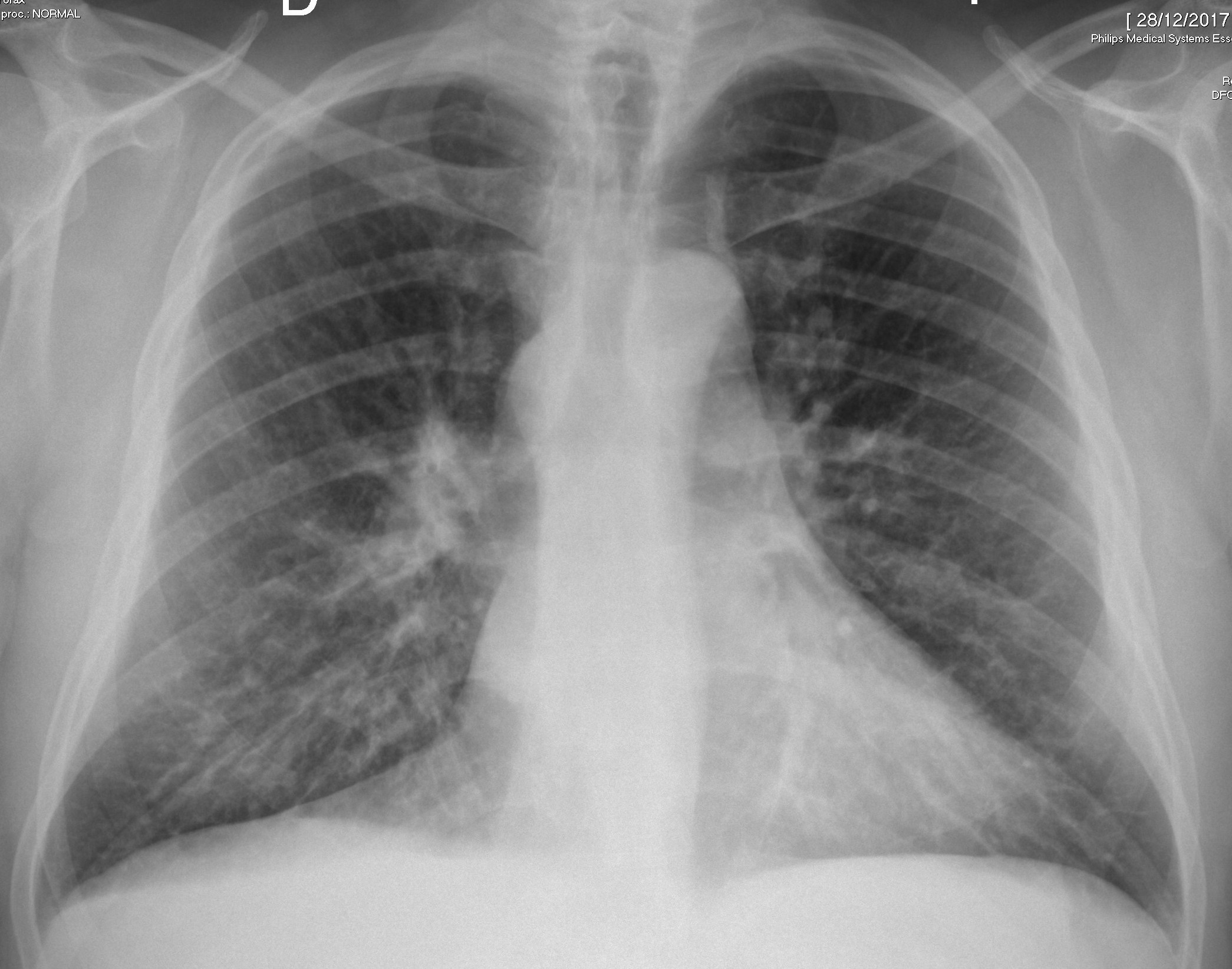
I want to present today a case that I saw two months ago. The radiographs belong to an asymptomatic 55-year-old man. Will show more images on Wednesday.
Check the images below, leave your thoughts in the comments section, and come back for the answer on Friday.
Diagnosis:
1. Enlarged azygos vein
2. Enlarged ymph node
3. Mediastinal mass
4. Any of the above
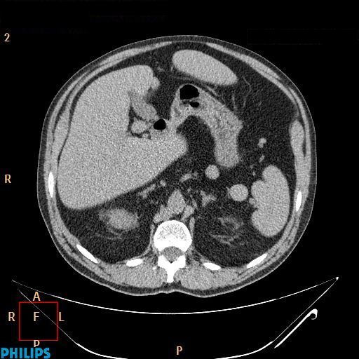
As promised, here are two additional CT images. What do you see?
Click here for the answer
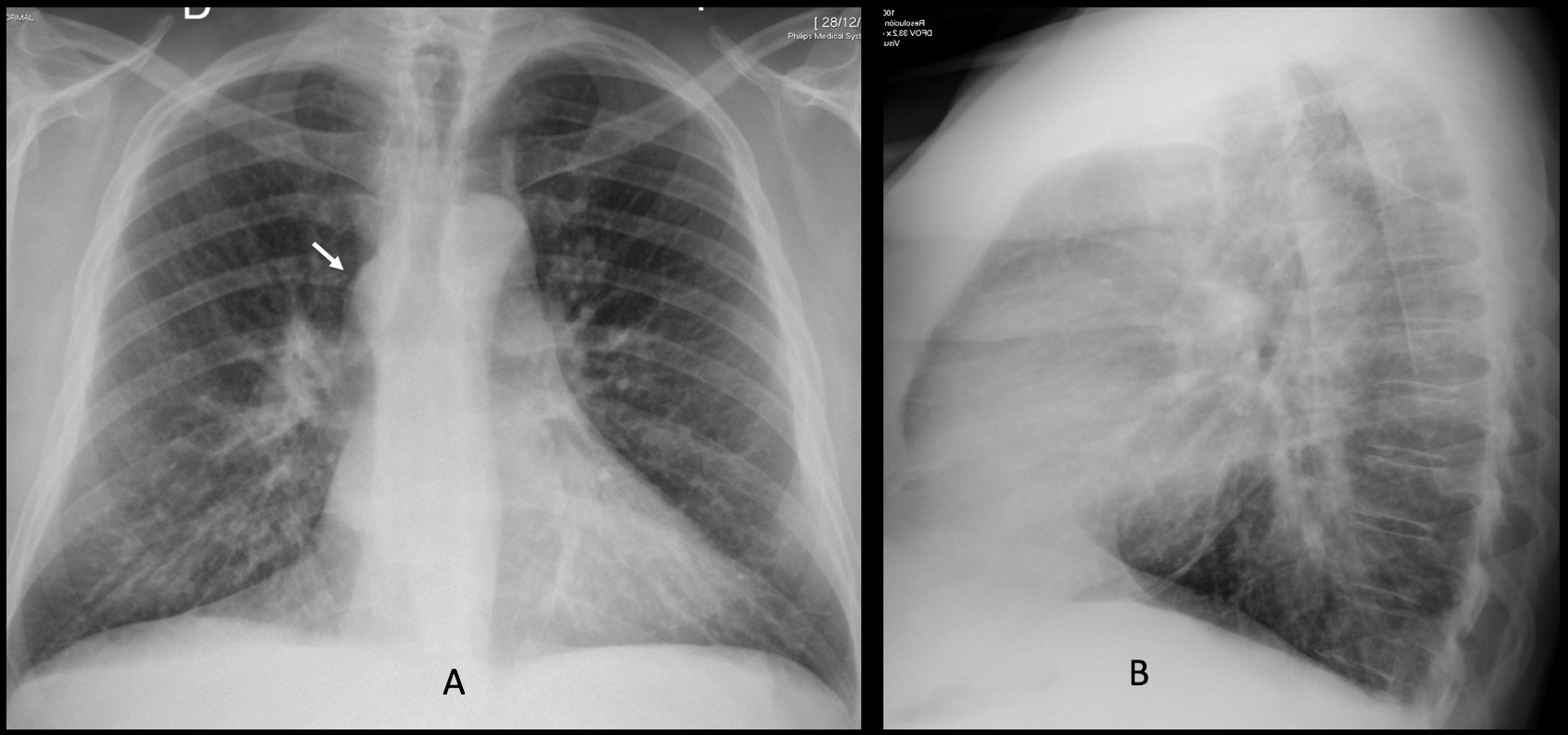
Findings: the PA radiograph shows a mediastinal mass at the level of the azygos vein (A, arrow), similar to the one seen in
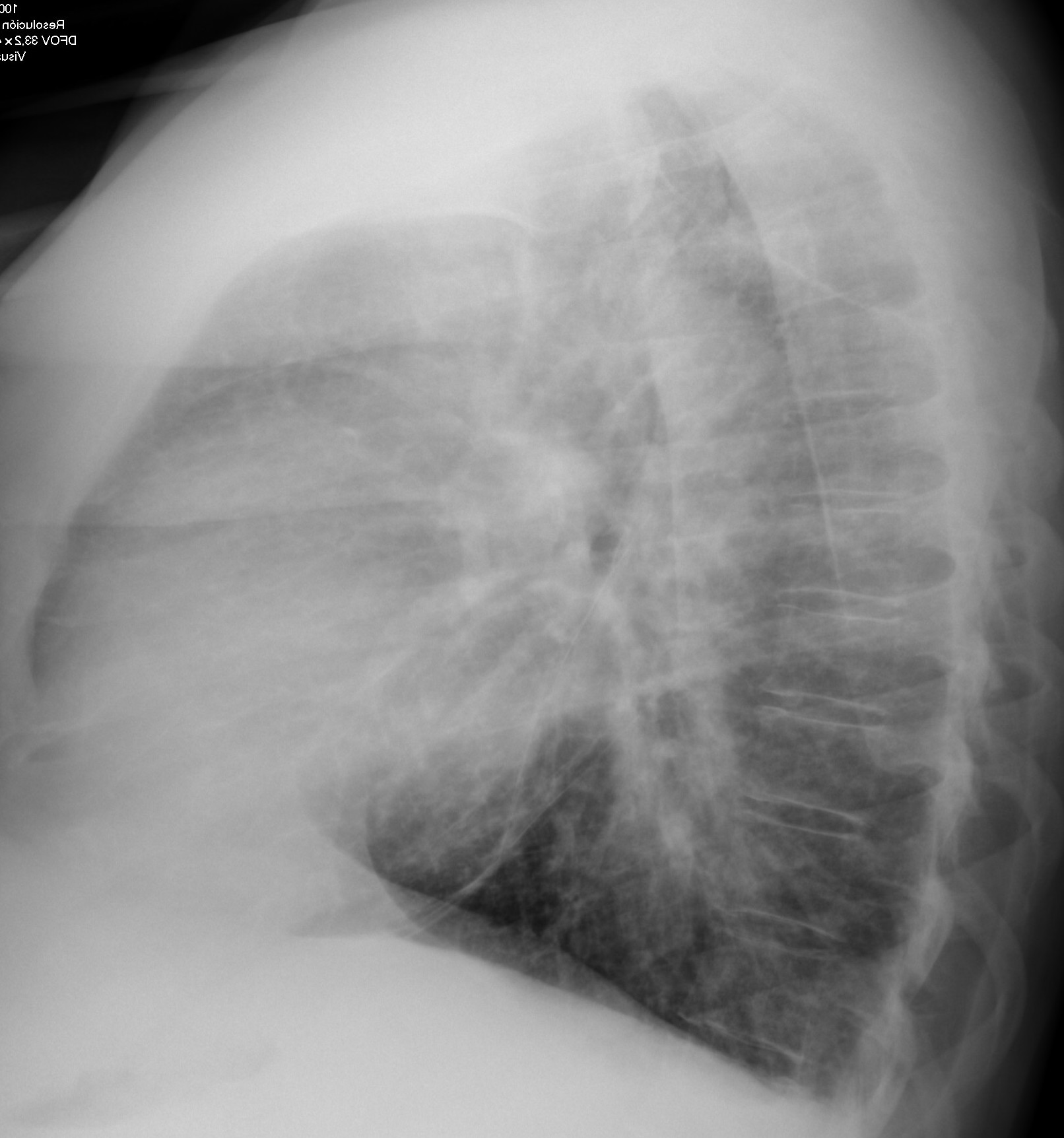
case 178. The same differential diagnosis applies. The lateral view (B) is unremarkable.
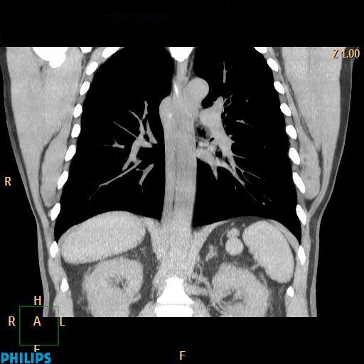
Unenhanced coronal and axial CT show an enlarged azygos vein (C-D, arrows). The inferior vena cava is absent. There are two nodules in the left upper quadrant of the abdomen (B-C, red arrows).
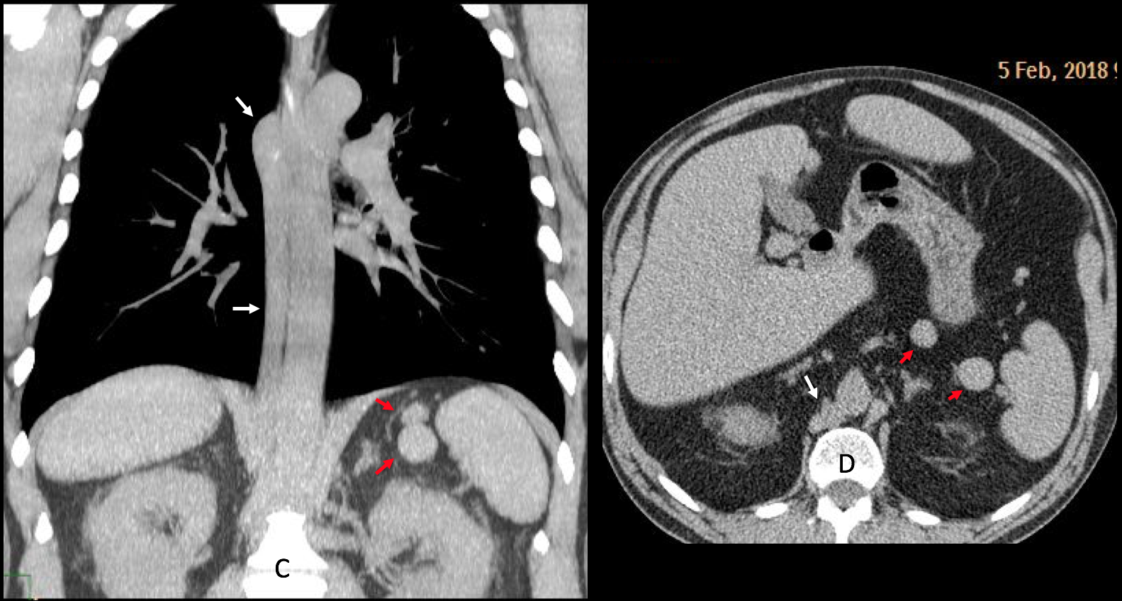
The findings are characteristic of a partially absent IVC which drains through the azygos vein. The nodules in the left upper quadrant represent accessory spleens.
I am showing this case to complement case 178, which had similar findings in the plain films that were not documented by CT. Also, both cases have been seen in a short period of time.
Final diagnosis: azygos continuation of IVC with polysplenia
Congratulations to MK, who was the first to suggest the diagnosis
Teaching point: congenital anomalies are not unusual. It is important to recognise them to avoid confusion with more serious conditions.


Good morning!
I think there is a right paratracheal lesion (in the lateral view it seems to be an adenopathy more than an enlarged acigos vein).
There is an spiculated lesion overlaying the right hilum.
I think we have to rule ot malignancy with a CT.
Chest radiograph AP view shows prominent and enlarged left hilum which is seen in lateral view also. There is paratracheal increased density seen on the left side just superior to the aortic knuckle, with continuation inferiorly and sail like traingular opacity seen upto left hemidiaphrgam. This is seen on lateral view as prominent oblique fissure.
It seems like left hilar lymphadenopathy with partial collapse left lower lobe. Suggest CECT chest.
Compensatory hypertrophy of the right lung is seen with herniation across the midline in anterior mediastinum.
Azygous vein also appears prominent.
Good evening Doctor Caceres! I have been following your teachings since 2015 and as a fifth year student in Brazil, I think I should start commenting on this blog instead of just being a silent reader.
Well, I think that there is an enlarged azygus arch to the right, opposed to the aortic knob, better seen in the lateral view. Also, I agree with MK about the spiculated lesion overlying the right hilum. Maybe an enlarged paratracheal lymph node? I think that these two lesions somewhat overlie each other in the lateral view.
Looking forward to the images on Wednesday!
Dear Renan, welcome to the blog! Would like to know your opinion about the new images.
Thanks for participating
Filling retrosternal space.
Right ventriculat heart configuration.
Congested lungs
My DDX are:
1 – lymphadenopathy, could be sarcoidosis (1-2-3 sign), right paratracheal and bilateral hilar LN enlargment (and the patient is asymtpmatic which corellates with stage 1 sarcoidosis)
2- right hilar mass.
Looking forward to see the diagnosis!
There is prominent density at rigth paratracheal region , possibly enlarged noe or prominent azygos vein.
Further CT show absent hepatic segment of IVC with azygos continuationof the IVC.
No right hiliar lesion.
Enlarged acigos vein with agenesia of the inferior vein cava with polisplenia, but it is difficult to confirm this with only 2 images.
You just did!
Intra tracheal
Enlarged acygos vein
This is Azygos as thers is no IVC.
Prominent azygous vien
Enlarged azygos vein