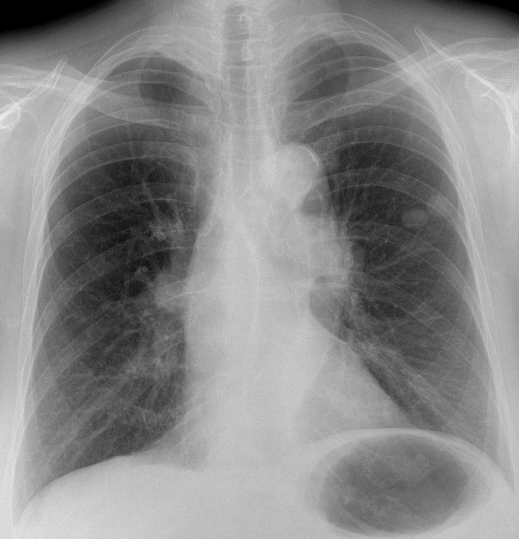
Today I am showing a pre-op chest radiograph for varices of a 79-year-old woman. A radiograph taken five years ago was normal.
What do you see?
Check the image below and leave your thoughts in the comments section. Will show more images on Wednesday and the answer on Friday.
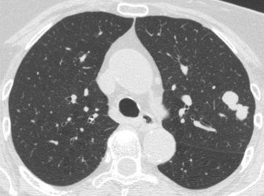
Findings: PA radiograph shows two nodules in the left middle lung field (A, arrow). In addition, there is a mass in the left hilum (A, red arrow). These findings were not present in a previous film taken five years earlier (B).

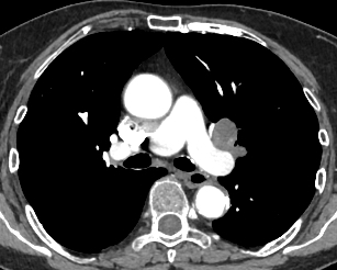
Enhanced axial CT shows lymph nodes in the left hilum (C, red arrows), as well as a dumbbell-shaped lesion in the left lung (D, arrow).
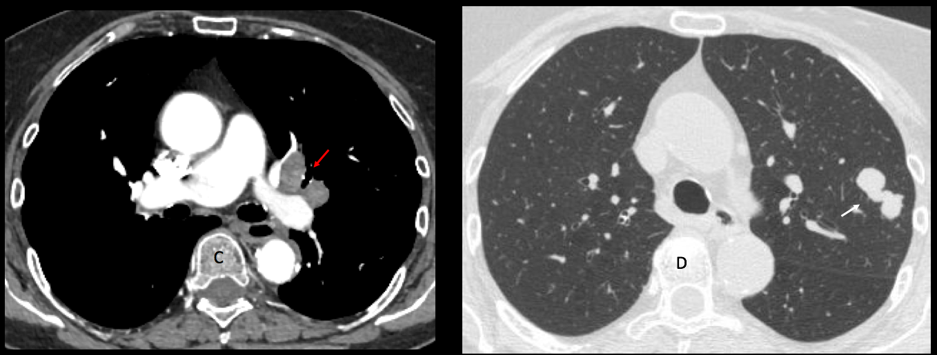
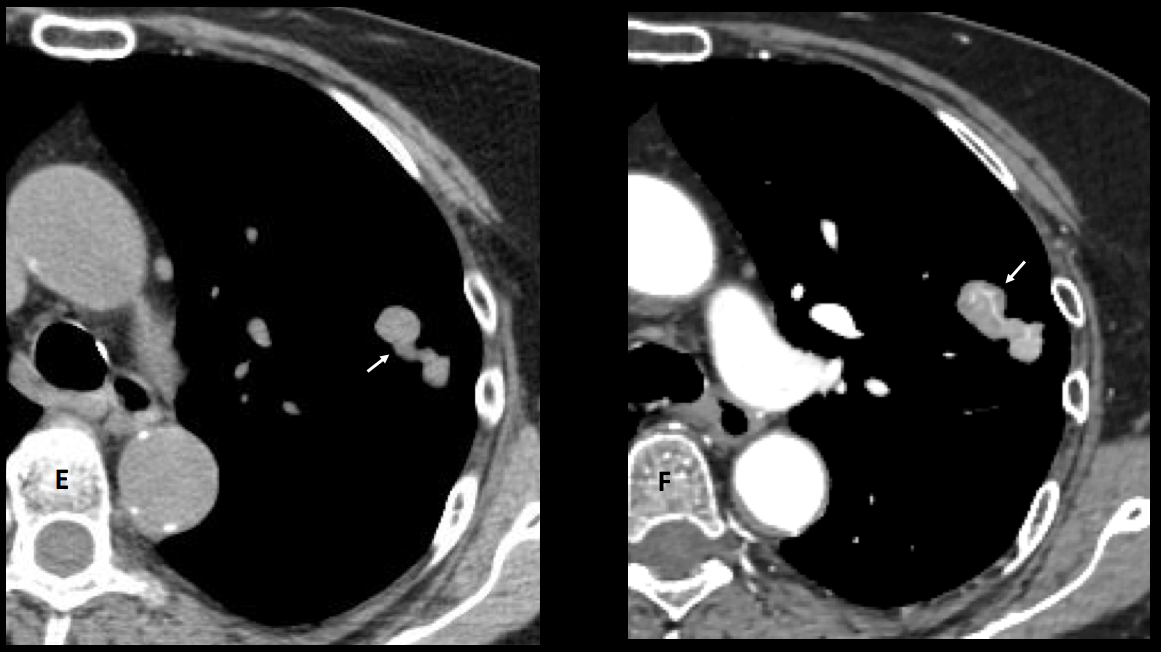
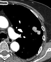
The lesion has meandering vessels within (E-F, arrows) and enhances 35 H.U. after contrast. A needle biopsy returned as small-cell carcinoma.
Final diagnosis: small-cell carcinoma of the lung with hilar metastases.
Congratulations to Trinity, who made an accurate diagnosis.
Teaching point: this case puzzled me. My original diagnosis was malignant fibrous tumour of the lung because of the odd shape and the presence of arterial vessels inside.
Unfortunately, reality defeated me. The lesson could be: always think of a rare manifestation of a common disease rather than a common manifestation of a rare disease. I stand corrected.


Hello sir,
There is increased density and size of both hila with loss of lateral convexity- bilateral hilar lymphadenopathy left more than right.
Two well defined round opacities are seen in LMZ-suggestive of pulmonary nodules- may represent metastatic nodules in the given age group.
The left hemidiaphrgam is also elevated more than right.
Linear reticular opacities in LLZ are seen parallel to left hemidiaphrgam, may be lymphangitis carcinomatosis.
My differential would be
1. Ca lung with satellite nodules and hilar lymphadenopathy.
2. Mediastinal lymphadenopathy with pulmonary mets.
Left hemidiaphrgam elevated due to mediastinal invasion and phrenic nerve palsy. Collapse of left lung not seen.
Hello Trinity,
Your answer and analysis make sense to me. But what do you think about the right pulmonary apex?
Best regards.
Thank you sir for your comment . Agree with your observation of right apical opacity, may be pleural thickening. Regards.
Apical opacity is not significant in this case. Look at the new images posted today.
Hello!
Prominents nodular hilar (adenopathy).
Two high density nodular lesions in left hemithorax, but I would like to see the lateral view to confirm that they are inside the lungs…
There is a lineal high density above the right hilum …?
Elongated aorta
Infection/fungal lesions??
Actinomices?
Bilateral hilar masses with two lt mid zonal large nodules and mild elevation of lt hemidiaphragm
DD
Lymphoma
Mets
Sarcoidosis
CECT scan shows left hilar mass and heterogenously enhancing pulmonary nodules left upper lobe. The right hilum appears normal.
Left hilar mass may be carcinoma lung with mets to LUL .
Thank you sir. Regards.