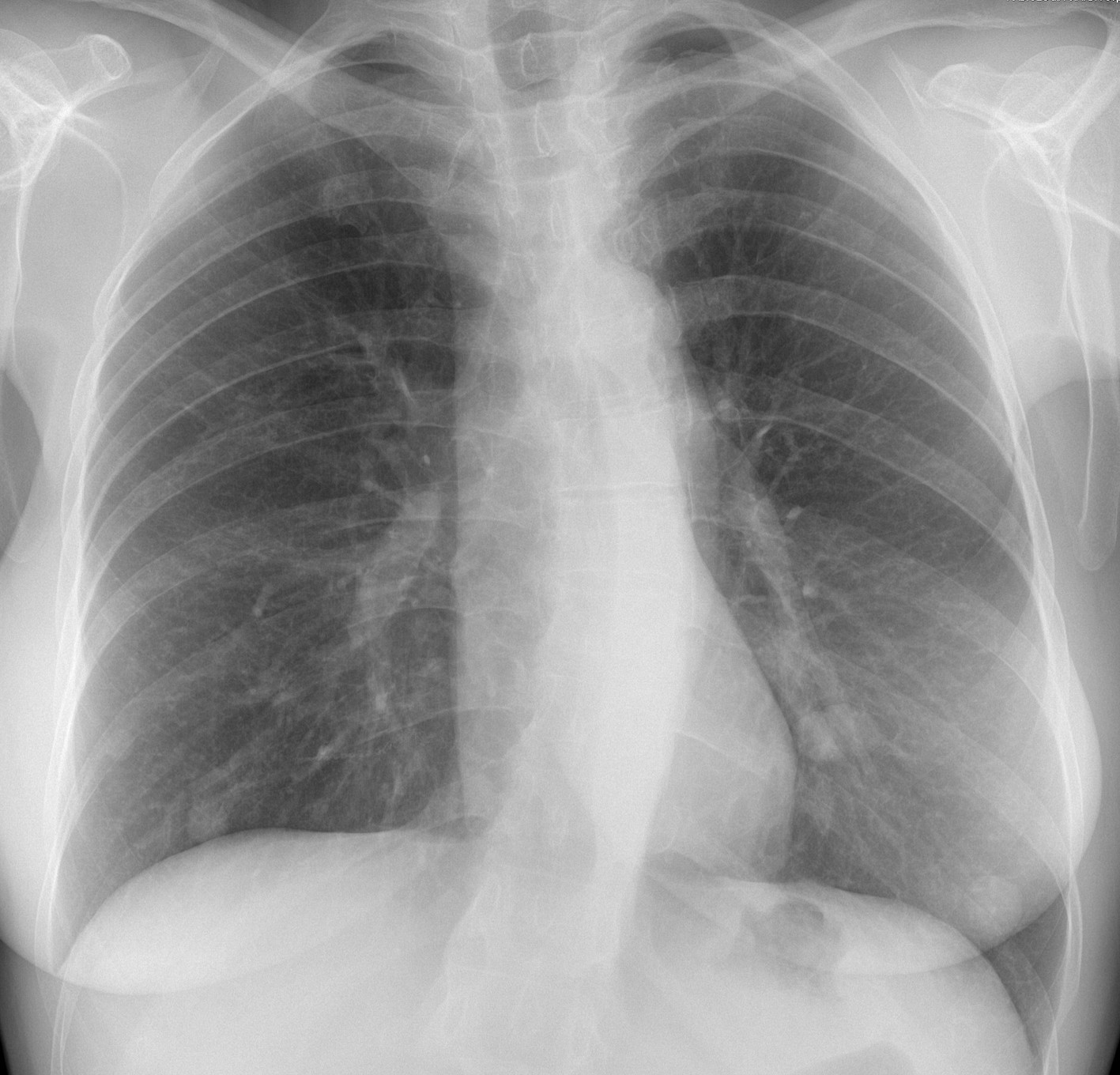
Today we are presenting a pre-op chest radiograph for knee surgery of a 48-year-old woman. What do you see?
Check the image below and leave your thoughts in the comments section. More images will be shown on Wednesday, and the final answer on Friday.
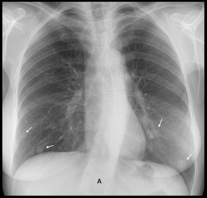
Findings: PA chest shows several nodules of unequal size in both lung bases
(A, arrows).
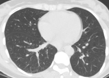
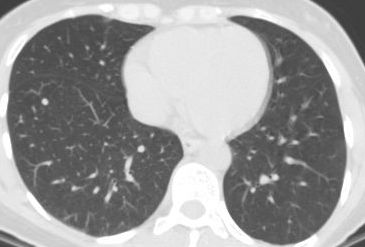
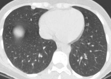
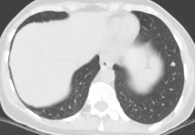
CT taken five years earlier for a study of hypertension demonstrates the same nodules, albeit smaller (B-E, arrows).

The differential diagnosis of slow-growing lung nodules is ample. The location in the lower lungs suggests haematogenous seeding and, in a female patient, metastasising leiomyoma should be considered. This particular patient had a history of uterine leiomyomas resected. At the time of the CT, a 7mm nodule in the RML was resected and diagnosed as leiomyoma.
At the present time the patient (a physician) refuses a new CT and/or needle biopsy.
Final diagnosis: Benign metastasising leiomyoma of lung.
Congratulations to all of you who faced a difficult case with dignity.
Kudos to Marco S, who suggested the correct diagnosis.
Teaching point: clinical history, location of the disease and previous studies are of great help in suggesting the right diagnosis.


There is a pulmonary nodule in the left lower lobe with well-defined margins. Maybe an AVM?
Marked scoliosis of the thoracic spine.
Only one?
of course there are more nodules 🙂 my mistake
No problem! What do you think?
Maybe hamartomas or AVMs. A CT (+/- CE dependent on the NECT) would be great 🙂 I can’t see any calcifications (burning calcification?)
Pulmonary AVM
Thorax PA film
-increased bronchovascular pattern (vascular: arterial origin)
-1 nodule in upper right lobe, 2 nodules in the lower right lobe,1 in the left upper lobe, and 1 more at left lower lobe near the left costophrenic angle
-Thoracic Scoliosis with left convexity
Otherwise everything looks completely normal to my n00b eyes ^^
Multiple -two in RLZ and three in LLZ- well defined lesions with smooth margins are seen in2 BLZ. No calcification or cavitation seen within these lesions. Being in periphery and basal lung fields, multiple pulm modules may represent,
1. Mets.
2. Rheumatoid Lung nodules.
Asymptomatic patient, no known primary.
The right cardiac silhouette and right paratracheal region show increased density with loss of normal contour. Is it rotation?
Probably due to the scoliosis. Don’t worry about it. More information tomorrow.
Multiple well defined lesions with smooth margins are seen in Bilateral LZ with no calcification or cavitation .
Levoscoliosis
Aortic calcification is displaced medially. Aortic dissection needs to be ruled out.
Dd
Mets
Rheumatoid nodules
Neurofibomatosis
Multiple bilateral well defined nodules..hamartomas?
Nothing on skin?
May be Nipples for two of them
No skin lesions. They are not nipples 🙂
Multiple well defined nodules at both lungs
Smooth regular margin for d.d
Hamartoma
Mets
Rh lung nodules
Maybe multiple fibroadenomas of breast. Pkus escoliosis.
In my experience, fibroadenomas of the breast are not seen in the chest radiographs unless they are calcified 🙂
Bilateral multiple nodules, right border of the heart is not seen, is it a pectus excavatum associated?
Pectus causes blurring of the right heart border and, sometimes, apparent cardiomegaly. It has no relationship to pulmonary nodules. And in this case I believe the right heart border is visible 🙂
I see 4 nodules in basal parts of lungs, with well-defined margins. Scoliosis. Of course, bilateral multiple nodules – “o, metastases” – shablon and first thought. But it can be granulomas. What is the cause of knee surgery?
Surgery was scheduled for a torn meniscus. No relationship to the nodules
Bilateral multiple nodules
And left side 3 4 5 bifid rib anomalies
Neurofibromatosi tipo 1: neurofibromi cutanei e scoliosi.Saluti carissimi PROF.
Well defined bilateral pulmonary nodules (are they slightly cavitated?) in a normal parenchimal.
Autoinmune process? (Wegener, reumathoid…)
Now you have all the information. Let’s summarize the findings:
Basal nodules discovered accidentally, slow-growing in an asymptomatic woman.
Think!
Pulmonary hamartoma fits the best.
There is a better alternative 🙂
lymphangioleiomyomatosis
Sarcoidosis
Do you mean leiomyomatosis?
Sorry, I meant leiomyomatosis. Multiple leiomyomas
Bingo! Congratulations
What a very interesting case! Thank You Prof. Caceres